So what is this all about? Well, there are always surgeries of the last-resort in any field.
As far as the eye, it is always a shame when only the cornea is messed up while the rest of the eye is intact. The logical next step is corneal transplant, typically around 90% success. What about the other 10%, though? They still need to see, don't they? Luckily, there are always surgeons who are willing to tinker, explore, invent, experiment, and cure. We will cite a few examples in this post.
Before we did that, a quick review on what could cause the corneal grafts to fail. Not surprisingly, the major one is allograft rejection, followed by increased intraocular pressure, infection (excluding endophthalmitis), and ocular surface problems. Pre-existing conditions such as diabetes and glaucoma increase the endothelial cell loss hastening the demise of the transplanted cornea. And chemical burn and end-stage dry eye (the latter as part of, e.g., Stevens-Johnson syndrome) both involving ocular surface changes also are major factors in graft failure. Any of the above leads to undesirable outcome, even multiple corneal transplants would not take.
The alternative is then to implant optically active prosthetics directly into the cornea. And if the retina is functioning well, then the patients can regain functional vision often at 20/40 or better.
First example is the Boston Keratoprosthesis developed by Dr Claes Dohlman of Massachusetts Eye and Ear Infirmary in Boston (a schematic is shown below):
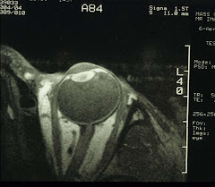
 The locking ring is titanium, it therefore does not interfere with MRI. This device is first fitted into a donor cornea, then the whole assembly is transplanted as that in normal penetrating keratoplasty. And a therapeutic contact lens is then fitted over the implant, together with life-long use of antibiotic eyedrops. The outcome is quite dramatic, interested readers can look up a news article "No time for tears" in the 5 Nov 2007 issue of Boston Globe.
The post-op appearance (without the contact lens) is shown below:
 The second example is OOKP (osteo-odonto keratoprosthesis) developed by the late Dr GianCarlo Falcinelli of San Camillo Hospital in Rome. In patients with severe dry eyes, Boston Keratoprosthesis may not perform well owing to the need for a contact lens - hence the need for adequate tear fluid production. These patients will have to have an eye-tooth implant. Eye-tooth = the upper canine tooth because it situates near the eye. Only a few surgeons in the world are qualified to perform this procedure that include Drs Christopher Liu of Brighton, England, Günther Grabner in Salzburg, Austria, and Konrad Hille of Homburg/Saar, Germany. And a team led by Dr Donald Tan of Singapore National Eye Centre is also active in OOKP implantation. Other eye centers may have also followed suit, check your local listing.
OOKP implant is a two-stage process. The first involves the repair of ocular surfaces using mucosal linings from the patient's cheek, removal of a canine tooth with part of the jaw bone, the tooth is fashioned into a bolt-shaped structure or a flat lamina with a hole drilled in the center, an optical cylinder is then inserted and cemented in the hole. The whole assembly is then implanted in the patient's cheek or under the fellow eye to allow growth of blood vessels. The second stage involves removal of anterior ocular contents and finally replacement with the tooth-bone-cylinder complex, now the tooth-eye.
Not all patients are surgically eligible for the procedures described above. When done, these last-ditch efforts often yield miracle-like results. These surgeons are a special lot, so are the patients who often have already endured multiple surgeries.


