With baby-boomers now entering the retirement age, there is an even greater need for geriatric care. We often hear from patients: "It is tough getting old..." However, short of providing water from the fabled Fountain of Youth, what we can offer is to try to maintain and repair.
The next series (2.X) will discuss various eye diseases associated with aging. And what kind of vision is expected and how to optimize/correct it. More for a comprehensive understanding from a researcher's point of view.
If you look at the tissues in the eye: in some, the cells renew quickly, while in others, the same cells are destined to last a lifetime. It is in the latter group, we have the age-related issue. Now a quick tour of the anatomy of the eye (diagram below courtesy of National Eye Institute, NIH, Bethesda, MD):
The cornea is bathed in tear fluid in the front and the aqueous humor in the back. Outermost is the epithelium with several layers that regenerate readily. All cells are replaced every 5-7 days. So any injuries to the epithelium is repaired quite nicely. The thickest middle is the stroma composed of collagen fibers that are maintained/repaired by keratocytes. And the innermost is the single-cell-layer endothelium whose cells do not divide if at all. With aging, the endothelium will lose cells. The endothelium can be regarded as a water pump which helps keep the cornea water content constant. The loss of cells by itself is not a major concern; however, additional losses from, e.g., corneal or cataract surgery or mechanical trauma from the IOLs can be a problem. The aging cornea is generally less resistant to infection, probably because of structural changes that have altered its barrier function. The periphery sometimes develop a white ring (known as arcus senilis) that can scatter light and reduce corneal transparency.
Next is the crystalline lens. It faces the aqueous humor in the front and the vitreous in the back. The adult lens structure, from surface to center is the cortex, adult nucleus, juvenile nucleus, fetal nucleus, and embryonic nucleus. And at the front is the single-cell-layer epithelium. The nucleus of course does not rejuvenate. Most of it is as old as your chronological age. The epithelium does go on producing cortical cell fibers which are layered upon older fibers. And the whole cortex-nucleus complex compacted with time (or you end up with age-related myopia which in fact does not exist). Throughout life, the lens is asked to refract light and itself will have received light-induced damages.
Finally, the retina which, as part of the central nervous system, never regenerates. Any retinal damage is therefore irreversible.
So now we know which tissues may be in trouble once age-related wear-and-tear kicks in and causes damages.
Saturday, December 1, 2007
Friday, November 30, 2007
1.9 What is a diopter?
Since there have been references to refractive power in the previous posts, a quick clarification is in order:
The diopter (D) is the basic measurement unit of optics; one diopter is equivalent to 1-meter focal length. The calculation of lens focusing power D is:
D = 1/focal length (in meters).
The higher the dioptric value, the stronger the lens focusing power; for example, a 10D lens will have a focal length of 0.1m or 10 times shorter than that of a 1D lens.
A plus (convex) lens has a real focal point (parallel light rays get converged onto one point) whereas a negative (concave) lens, a virtual focal point.
In the eye, the corneal power is about 44D (taking into account the refractive index of air=1 and that of the aqueous humor=1.33), and the power of the crystalline lens about 16D - for a total of 60D. This is essentially a 15X magnifier (60D/4 = 15X assuming 25cm of viewing distance).
Incidentally, if the crystalline lens is removed as in cataract surgery, then all 16D must be restored using spectacles, contacts, or more frequently intraocular lens implants (IOLs).
Only in lay terms, for example, -1.25D = 125 degrees of myopia; although there is really no such thing as degrees of power in optics.
Thursday, November 29, 2007
1.8 Other kinds of myopia
In addition to school myopia, there are several other kinds of myopia, mostly secondary. To complete the discussion on myopia, I will also mention them.
Usually this type of myopia drives everybody nuts. Because the refractive error does not stablize, it is very difficult to know if the endpoint of refraction is reached. Often the patients complain. The opticians blame the doctors for "measurement errors". The doctors re-refract ending with minimal changes and try to explain the underlying factors to the patients, usually in vain. And the patients are still quite unhappy because they expect 20/20 vision with a new pair of expensive yet now apparently useless spectacles.
It is actually quite simple, any condition that increases the overall refractive power of the eye will lead to myopia, sometimes transient, often part of permanent or semi-permanent ocular pathological changes. The possibilities are (1) increase in the refractive index in the nucleus of the crystalline lens; (2) steepening of corneal curvature and/or corneal edema; (3) sustained accommodation - prolonged accommodation precipitates pseudomyopia which usually recover once the stress factor is removed; (4) medicine-induced: drugs such as sulphonamides can cause acute transient myopia; (5) ocular surgery, e.g., retinal detachment repair in which scleral buckling around the eye globe can deform the eye and lengthen the axial length; (6) tumor-related increase in the vitreous volume or the anterior-posterior length of the eye, even though intraocular and intraorbital tumors are rare.
Most commonly encountered are actually (1) and (2):
(1) Lenticular change: The refractive index of the nucleus can increase as a result of aging or diabetes, often part of nuclear sclerosis that eventually leads to nuclear opacities (i.e., cataracts). Diabetes actually causes more than just lenticular changes (more when we discuss cataracts). In both cases, myopia induced by nuclear cataracts frequently occur. The rate of myopia increase is, however, unpredictable. This type of myopia is often accompanied by deteriorating vision.
(2) Corneal change: Typically long-term contact lens wear can result in corneal warpage. And chronic oxygen deprivation can result in corneal edema with changes in both thickness and refractive index. Again, these manifest as gradual myopic increase throughout the contact lens wear period, known as the "myopia creep". If the patients stop wearing contacts, or switch to high-oxygen transmission lenses, the corneas tend to return to the baseline albeit slowly. This type of change is also a major concern prior to refractive surgery.
1.7 What about genes?
Even now, we are still blitzed by the media as far as "gene of the week", yet so far no "breaking news" on the myopia front. No, it is not as if no one is interested. Quite the contrary. It is just that nobody knows if there is only one myopia causative gene or an assortment of genes working in concert or in tandem, and what the gene products might be. Or, horrors, maybe there is no such thing as a school myopia causative gene at all. Whatever it is, it is a daunting fishing expedition involving several modes:
Matter-of-fact mode: "Our work confirms a previously reported [refractive error] linkage region on 22q and identifies 2 novel regions of linkage on 1q and 7p. Further, genetic research is needed to finemap this trait to identify the causative gene" (Beaver Dam Eye Study).
Exclusion mode: For example, one report points out that neither PAX6 nor SOX2 should be a priority in the search for genetic modifiers of myopia, and in another: sequence variants of the TGF-beta1 gene are not associated with high myopia.
Inclusion mode: For example, in an Australian study, axial length is found to be highly heritable and is likely to be influenced by one or more genes on the long arm of chromosome 5.
Humpty-Dumpty mode: hereditary high myopia loci (MYP12) may contribute to all degrees of myopia.
And to make it even more challenging or muddy-the-water mode: a study finds that an X-linked high myopia locus is mapped to Xq25-q27.2 which overlaps with that of MYP13 but is outside MYP1.
These are just a small sample of reports published in 2007, from all over the world. You can go to http://www.ncbi.nlm.nih.gov/sites/entrez and enter keywords: "gene and myopia" to find the above and more. Notice, however, that they are not genetics of school myopia per se. The emphasis is still necessarily on families with high myopia which is usually not morphometrically defined. So we don't know if the eyes contain a posterior staphyloma component.
Ideally of course, if a causative gene can be identified, then we are talking gene therapy to decrease the risk of developing myopia. The other side of the coin is, what are the environmental factors that initiate or contribute to myopia gene expression. Then modifying these factors is probably as effective as the genetic approach.
Ah, now we are back to nature or nurture.
In a paper published in 2005, a mutation in MFRP gene is linked to nanophthalmos (dwarf eye) which is an under-developed, very hyperopic eye. And the missing MFRP protein has been proposed to regulate the eye growth. Very intriguing finding indeed.
You know what I think? Since myopization involves expansion of the eye globe implying a re-structuring of collagen fibers in the sclera. Then it is probably one of the MMPs that is asked to loosen the inter-fiber bonds - through the mediation of the MFRP protein. By concentrating on this one aspect, perhaps this MMP can be isolated and an inhibitor developed. Nothing fancy, just old-fashioned biochemistry. Much like using the statins to inhibit HMG-CoA reductase and stop cholesterol synthesis all together.
1.6 Contacts or LASIK?
For those with school myopia, there are always alternatives to spectacles. You know the old sales pitch: you see better/more with the contacts; with LASIK, you can see the digital clock clearly in the middle of the night... It maybe worthwhile to go into contact lenses and refractive surgery as indeed these are not your grandfather's limited choices anymore.
Contact lens is a marvel of modern technology. It is small, from 9-15mm in diameter, and light-weighted (less than 0.02mg) that can be worn comfortably for clear vision. However, it is still a challenge to corneal physiology that is based on aerobic glucose metabolism. Biochemistry 101 will have told you that in aerobic glycolysis, 1 mole of glucose produces 38 moles of ATP, while in anaerobic glycolysis, only 2 (plus lactate). A large part of contact lens R&D is devoted to the search for materials that allow high oxygen transmission. And Voilà! The most recent generation of contacts combine water and silicone (the silicone hygrogel lenses) now permit 5-6 times more oxygen than the older yet still popular HEMA-based soft lenses.
Of course, not all kinks have been ironed out. Contact lens is simply a device. Within the framework of contact lens wear, we still need to ensure that the cornea remains reasonably healthy, totally free from immunological and microbiological issues. Sometimes, the problems are unexpected. A case in point:
In the summer of 2005, the Health Department of Hong Kong was alerted to cases of Fusarium solani fungal keratitis. It is a devastating infection that often leads to the destruction of the cornea. The Ministry of Health of Singapore then also reported that between March, 2005 and May, 2006, 66 cases (68 eyes) were diagnosed, 65 wore soft contact lenses and 62 used Bausch & Lomb ReNu (42 on ReNu MoistureLoc) solution. The first case in the US was reported on March 3, 2006, and subsequently, 164 cases in all were identified. Of which, 124 cases reported using ReNu MoistureLoc. On April 13, this solution was removed from the US market, and on May 15, from the World market. Exhaustive investigation, however, failed to uncover any deficiencies in the manufacturing process. The "Fusarium epidemic" on the other hand, was over. The suspicion is that some polymer components in the solution, when dried on the surface of the contact lens case or the tip of the solution bottle, may have harbored this fungus.
Let's not forget hard contacts, now highly gas-permeable that allow 100 times more oxygen than the soft lenses. And relevant to the myopia discussion is the use of GP lenses to retard myopia progression. Thus far, the evidence points to a limited effect similar to that in multifocal clinical study mentioned in "Children with bifocals" (Nov 28, 2007). And there is always an element of orthokeratology.
A little more on orthokeratology: it is popular in some parts of the world/US. This procedure takes advantage of the deformability of the cornea. By sleeping with gas-permeable hard contacts with the back surface curvature flatter than the cornea, the corneas will stay flattened for a few hours sometimes longer during daytime. Clear vision is thus achieved, seemingly without optical aids. This ortho-K effect is not permanent, though. And wearing contacts during sleep has some inherent risks. The eyes may become inflamed/infected and the lenses may adhere to the corneas necessitating an emergency visit to the doctor's office. All part of the deal.
If you wear contacts, soft or hard, ortho-K or otherwise, do follow the doctor's instructions. Of course, it is also possible to chuck the contacts all together and go straight for refractive surgery.
For myopia surgery, it is a matter of how to flatten the corneal curvature to remove power from the optical system of the eye. This was first done by the now out-of-favor RK - with radial cuts on the cornea but sparing the pupillary area. After healing, the central corneal curvature flattens. Using the Excimer laser to blast off tissue and reshape the cornea is the standard these days. We now have PRK and LASIK. LASIK (and the like) has one extra step: lifting the corneal epithelium before applying the laser. The epithelium is re-attached after laser so the healing is faster and far less painful than PRK.
A pre-op consultation will first make sure your corneas are healthy, refractive error is stable, corneal contours are regular, and most important, the corneas are thick enough. With the advent of wave-front technology, it is possible to maximally improve your vision. If high myopia with thin corneas, the alternative is to implant an intraocular lens (i.e., a phakic IOL).
As in all surgery, refractive surgery carries certain risks; although none involves irreversible vision loss - none reported in the US thus far. And the complications can be severe dry eye, halo or glare at night, or over-/under-correction. These become less frequent as both laser technology (now wavefront-guided) and surgical procedures (e.g., new ways of creating "flaps") have vastly improved.
Contacts or LASIK? For young and healthy eyes, either one is good. For middle-aged people, first think about how close you are to wearing reading glasses. It is now only a matter of choice; doctors can take care of the rest, usually.
Wednesday, November 28, 2007
1.5 Children with bifocals
Before the medical prevention/treatment of myopia becomes a reality and a safe one we hope, we are still stuck with glasses, contacts, or refractive surgery.
Now, if we assume there is a role for accommodation in myopization, then is it possible to use spectacles, instead of cycloplegics, to prevent myopic progression? Yep, it has been done before. And the results apparently were not all that spectacular or you and I would have been wearing bifocals since what, middle school? The problem seems that children with bifocals have a way of peering over the bifocal part to read, plus they lose some navigational agility when, for example, rushing downstairs or playing soccer. The data are highly suspect if the children did not wear those bifocals full time. So a recent clinical study chooses progressive bifocals, i.e., multifocals instead. And the data are far more reliable because of the superb experimental design and diligent follow-ups. The results are a little encouraging or discouraging, depending on your perspective. The bifocaled children in the study do show a smaller increase in myopia comparing with those wearing single-vision spectacles. The difference is, however, less than 0.25D and only during the first year. This therefore does not warrant a change in clinical practice in managing school myopia. On the other hand, "defocus" does have a role in myopia progression. So it is back to chicken or egg first. What in the world triggered this defocussing?
Because of this persisting and may even be correct notion that accommodation is the basis of myopization, over the years, there have been different strategies, e.g., under-correction (common practice in Asia), read without spectacles, etc. Mostly for parents' psychological benefits, though.
The math of accommodation is quite simple. For reading at 13 inches (i.e., 40 cm or 0.4 m), you'll need to focus 1/0.4 = 2.50D and so on. You start out as a child with a full accommodative power of around 14-16D which decreases with age pretty much linearly until about 0.25D when you are 70 years old. It depends on how much accommodative reserve your have left. Let's say 1.50D, then you will need either longer arms to hold your newspaper at (1/1.5=) 67 cm, or another 1D in the form of reading glasses to make up the difference - in order to read at 40 cm. By the same token, if 2.50D is to be eliminated from children's near focusing, then 2.50D is provided for in those bifocal/multifocal clinical trials. And if 2.50D is needed for reading at 40 cm, why read at 20 cm as some children do, with (1/0.2 =) 5D, or 5 - 2.50 = 2.50D extra, unnecessary accommodation for reading the same Harry Potter thing.
As far as myopia prevention, back to the drawing board.
1.4 Jumping the gun
Of course there are always innovative doctors, some have tried to medically "cure" myopia or prevent myopia from progressing. Again, this dates back several decades. The rationale was since near work, i.e., reading (this was the pre-CRT ancient period, no computers or TVs, what video games, etc) seems to cause myopia, and since reading requires accommodation, then by paralyzing accommodation, myopization should be preventable.
Accommodation is a process by which the crystalline lens inside the eye increases its thickness and curvatures through the contraction of the ciliary muscle. This way more refractive power is gained for focusing at close range. Incidentally, in people over age 42, the crystalline lens can no longer change shape. Reading glasses are therefore needed to supplement the power. This condition is known as presbyopia. (Note: If you have distance correction as well, then get multifocals; when made correctly, they are wonderful.)
Funny thing is that by using a group of cycleplegic eyedrops which not only dilate your pupils but also shut down accommodation, myopia progression was actually slowed and sometimes reversed in treated school children. Great news, right? Well, not quite.
The problems are (1) pupils remain dilated for long periods of time, meaning sunglasses are needed and (2) no accommodation, no reading. Neither is compatible with the lifestyle of a school child. I would have chosen to wear glasses myself. Plus, no one knows the long-term effect of these cycloplegics.
So the medical treatment has been semi-forgotten for a while until more recently when there has been some change in the dosing regimen. For example, the cycloplegics can be applied to only the more near-sighted eye until the other eye catches up with the refractive error, then treat the other eye next, and so on. This type of monocular mydriasis/cycloplegia is more acceptable and well-tolerated by the children. Ideally, if only accommodation is paralyzed but the pupil dilation is spared, i.e., more specific eyedrops. Then the use of these drops should be promoted, especially for prevention. Let's say, 80% of the school children will eventually develop myopia, then by treating all, we are only over-treating 20%. Not too bad a deal.
Even more recently, there have been trials of accommodation-specific eyedrops. Unfortunately, the results are not that much different from the old cycloplegics.
Now, is the cycoplegic treatment of myopia based on irrefutable scientific evidence? Not really. We just know the eyedrops worked. Obviously more R&D is needed.
Come to think of it, the market size for this type of eyedrops is actually quite mind-boggling; although the optical industry may suffer recession as a result. You win some, lose some.
A word of caution: All cycloplegics for myopia are still in clinical trials done only under doctors' direction and care. It is not a DIY project or you'd be suing yourself for practicing medicine without a license or worse, being sued for child abuse.
Tuesday, November 27, 2007
1.3 Myopization
In order to study myopia formation or myopization in the lab, animal models are a must.
The development of animal models of myopia dates back several decades. The earliest attempt had monkeys' heads enclosed in a small box, so they could not see far away. This was to simulate close work, or more accurately induce sustained accommodation. And indeed, these poor monkeys did develop some myopia. One look at an active human child, however, you know immediately that this monkey-myopia is probably not a good model - even though the basic biochemical mechanisms may be the same. This can be very important (see below).
More recently, deprivation myopia models have become very popular. For example, if one eye of a chick is occluded with a translucent cover for a period of time, that eye will become myopic. You can do the same to monkeys, mice, guinea pigs, tree shrews, etc, and get the same result. In fact, in human babies with monocular congenital cataracts, that eye becomes myopic as well. In other words, "not seeing" can cause uncontrolled eye growth resulting in myopia. Along the same vein, by using contact lenses of varying power with/without cylinders, all kinds of refractive errors can be duplicated. So "not seeing well" can be an inducing factor. And if you cut the optic nerve but maintain the viability of the eye. The eye develops myopia also. Then "not seeing at all (blindness)" is yet another factor.
On the other hand, school myopia starts out with normal vision that gradually evolves into myopia with no form deprivation, etc, at all. How do we reconcile the difference between animal models and human school myopia? Not very easily, I am afraid. However, this does not mean that animal models are useless. The saving grace is this: the fundamental biochemistry of myopization may still be common to both. Then our efforts should be directed at elucidating myopia biochemistry (see Myopia, Nov 26, 2007).
1.2 Myopia is a good thing?
Most if not all biological changes are for a good reason, i.e., they are not random processes. Development of myopia is no exception. A simplistic view is: Perhaps the collective human brain computes that when urbanized kids are growing up, they already need to work at close range, and the situation continues into the adulthood anyway, so it is advantageous for all to see clearly at near without the need for much accommodation. There may be some truth here. If you are a myope, take off your glasses/contacts, sit back, and look at the image below:
You can actually see more clearly the face of Mona Lisa. Isn't that interesting. The real da Vinci code! In other words, the edges of all the pixels are smoothed out. This maybe an advantage in the computer age?
Well, what about after you leave the office and try to drive home in the midst of a blizzard? And who's gonna fly the commercial/military airplanes or become your local firemen/policemen? Blurred distant vision is no longer so great, is it? Shouldn't the brain now tells the eyes to develop a different adjustable focusing mechanism as that in some cameras (the same lens moving in and out), just in case?
Perhaps the human brain/visual cortex is not all that smart. Or maybe it is: "Hey, not to worry, we have glasses, contacts, and LASIK..." Or as they say, evolution takes millions of years, if not longer. Yeah, right.
Current research is aimed at figuring out the myopization process and how to put a stop to it. There have been "progresses" on both fronts.
We'll touch upon the myopization process next.
Monday, November 26, 2007
1.1 Myopia
OK, we all know what myopia is. So what causes it? There is a Nobel Prize in there somewhere if you can come up with the answer. To be specific, we are talking about "school myopia", the type that starts up usually in elementary schools, not those induced by, e.g., cataracts or diabetes mellitus. Of course, there has been a long debate on whether it is nature or nurture. Which is probably irrelevant - if your eyeballs are predisposed to developing myopia, once given the stress, myopization will most likely ensue.
First, let us look at what we do and don't know:
1. Environmental factors: In survey after survey in Asian countries, myopia starts to show in elementary school children, by college years, most students are myopic. In some medical school classes, non-myopes are a rare species. In contrast, in rural areas of China, myopia is not as prevalent as that in the cities in school children of the same age. So now: Too much education (hint: too much reading) is a risk factor? Move medical schools to countryside?
2. Family tradition: If you look into your family photo albums, you'll notice no one wore glasses a few generations back. Not that the glasses were unavailable, mind you. They were. It is definitely in the more recent generations that myopia becomes rampant. A common scenario is a family with one or both grandparents myopic, so are the parents, and chances are the next generation will also be myopic. So what has had happened between the often non-myopic great grandparents (and even earlier generations) and the grandparents of the present time? Something in the water?
3. Mothers' concerns: You must have heard mothers telling their children not to watch too much TV, play video games, surf the net, etc, or risk becoming near-sighted? So what is the truth - excessive near work precipitates myopization? If so, then reading/studying should be banned as well?
Speaking of myopization, what are the processes? Perhaps we can take some educated guesses:
Well, since high intraocular pressure is not associated with school myopia, we can probably rule out glaucoma genes. Of course, some argue that all myopes are glaucoma-suspects until proven otherwise. This is, however, a later complication, not a primary causative factor.
Structurally speaking, myopic eyes have very thin sclera and thin choroid as well. This seems to imply that (1) a re-arrangement of collagen fibers of the sclera, in other words, the thin sclera is not a result of ballooning from high intraocular pressure ; (2) a reduction of the blood volume in the choroid; and/or (3) an increase in vitreous cell production of vitreous collagen/hayluronic acid, i.e., an increase in the volume of the vitreous. A closer look into the biochemical mechanisms of all three then makes sense. Perhaps the starting point should be posterior staphyloma that usually is the chief culprit of exceedingly high myopia, e.g., -10D or more.
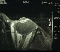
Posterior staphyloma is readily seen with either ultrasound B-scan or high-resolution orbital MRI. It is a small bulge out from the posterior pole. With A-scan, you'll be under the impression that the eye has long anterior-posterior axis. Current clinical studies of myopia progression are all based on A-scan which obviously cannot detect posterior staphyloma.
A quick note on the math of myopia: At roughly 1mm (along the anterior-posterior axis)=3D (diopter), you can see why myopia increases so quickly as 1D is equivalent to a mere 0.3mm, so an emmetropic eye is around 23mm "long", an eye with -10D refractive error will be 26.3mm "long" well tolerated within the orbit. Of course there must be an upper limit for the eye globe to fit into the orbit without the appearance of that in Graves Disease (i.e., exophthalmos). So an eye with -20D, or almost 28mm "long" will not move too freely when looking laterally. I have used long in quotes because this is a one-dimensional measurement. Myopic eyes are in fact huge in the 3-dimensional sense. Usually, an adult eyeball is 6.5ml and the orbit is 30ml in volume. Besides the eye, there are fat, blood vessels, the optic nerve, and the six extraocular muscles also. In Graves Disease, either the muscles or the orbital fats become bulky thereby forcing the eyeball outward.
Back to posterior staphyloma: Apparently some kind of signals have ordered an out-pouching of the posterior pole. Then we maybe looking at two different collagen fiber re-arrangements in the sclera, an early one for the school myopia and the later second for posterior staphyloma. And it should be a matter of identifying what these signals are. For example, growth factors or MMPs may have been dispatched in two waves by the emmetropization control center somewhere in the visual/cerebral cortex.
This brings up another question: is myopia a good thing?
Sunday, November 25, 2007
Greetings
I have decided to post my lecture notes as well as share my thoughts on some unresolved research issues. Some of you maybe interested enough to formulate research projects based on these topics. I will post them one at a time that will cover the anterior segment all through the visual pathway to the visual cortex. Since it is a work in progress, you may want to review older posts for new info. Feel free to post back, naturally.
Subscribe to:
Posts (Atom)