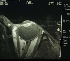
In order to study myopia formation or myopization in the lab, animal models are a must.
The development of animal models of myopia dates back several decades. The earliest attempt had monkeys' heads enclosed in a small box, so they could not see far away. This was to simulate close work, or more accurately induce sustained accommodation. And indeed, these poor monkeys did develop some myopia. One look at an active human child, however, you know immediately that this monkey-myopia is probably not a good model - even though the basic biochemical mechanisms may be the same. This can be very important (see below).
More recently, deprivation myopia models have become very popular. For example, if one eye of a chick is occluded with a translucent cover for a period of time, that eye will become myopic. You can do the same to monkeys, mice, guinea pigs, tree shrews, etc, and get the same result. In fact, in human babies with monocular congenital cataracts, that eye becomes myopic as well. In other words, "not seeing" can cause uncontrolled eye growth resulting in myopia. Along the same vein, by using contact lenses of varying power with/without cylinders, all kinds of refractive errors can be duplicated. So "not seeing well" can be an inducing factor. And if you cut the optic nerve but maintain the viability of the eye. The eye develops myopia also. Then "not seeing at all (blindness)" is yet another factor.
On the other hand, school myopia starts out with normal vision that gradually evolves into myopia with no form deprivation, etc, at all. How do we reconcile the difference between animal models and human school myopia? Not very easily, I am afraid. However, this does not mean that animal models are useless. The saving grace is this: the fundamental biochemistry of myopization may still be common to both. Then our efforts should be directed at elucidating myopia biochemistry (see Myopia, Nov 26, 2007).
Subscribe to:
Post Comments (Atom)


No comments:
Post a Comment