Saturday, December 15, 2007

5.4 People who need people

Barbara Streisand may have been describing epidemiologists, you think?

Epidemiology is a fascinating field of study. In fact, large-scale population studies are now relatively common. The conclusions are, however, confusing to the general public at times.

In 2001, the National Eye Institute sponsored AREDS (Age-Related Eye Disease Study) reported that nutritional supplements significantly reduced the progression of early AMD. These supplements included

  • 500 milligrams (mg) of vitamin C
  • 400 international units (IU) of vitamin E
  • 15 mg of beta-carotene
  • and 80 mg of zinc oxide
In 2004, however, news headlines screamed "Vitamin E can cause death", based on this study:

Miller ER 3rd, Pastor-Barriuso R, Dalal D, Riemersma RA, Appel LJ, Guallar E. "Meta-Analysis: High-Dosage Vitamin E Supplementation May Increase All-Cause Mortality." Ann Intern Med. 2004 Nov 10.

Some AMD patients who were on vitamin E supplements went into the panic mode, when in fact the "high-dose" in the 2004 report refers to >>400IU daily intake. The headline writers were somewhat irresponsible.

Ideally, all comparative epidemiology studies employ the same methodology so that the comparisons can be done. One good example is the cataract prevalence studies, most of which use their own home-versions of cataract classification. Your idea of a nuclear cataract-caused blindness may just be my 20/200 vision with a NS3+. So how are we going to compare the data? Eventually, the WHO organized a Cataract Grading Group who published "A Simplified Cataract Grading System" in the September, 2002 issue of Ophthalmic Epidemiology. However, to this day, the WHO system has not been widely adopted, let alone applied. Inertia is probably at the root.

It is really just common sense. For international studies, a common methodology must be agreed upon and strictly adhere to. The results will be far more useful for worldwide public health planning.

5.3 Here comes the sun

The Beatles may have gotten it partially wrong. Here comes the sun, but it is not quite all right, as the sun can do some serious damages to your eyes. And we are talking radiation from ultraviolet (UV) which has two components, UVA: 280–320 nm and UVB: 320–400 nm. (Hint: when purchasing sunglasses, make sure they block both UVA and UVB.)

Sun/UV damage to the eye comes in many flavors. The one we still occasionally see is solar retinitis, i.e., staring at the sun with unprotected eyes for a prolonged period of time. The reason for this behavior is unknown, it certainly is a good way of burning up your fovea quickly, resulting a small central scotoma. Sometimes there is recovery from re-grouping of the cones. Most often, not.

Then there is "sunburn" of the cornea, i.e., solar/UV keratitis. The source of the UV is not necessarily the sun, it can be a sunlamp or welding arcs. Needless to say, it is quite uncomfortable.

The most prevalent UV-related damage is actually pterygium.

Pterygium is a benign fibroblast growth from the conjunctiva into the cornea (the more localized form is called pinguecula). The image below shows a mild case. Occasionally it becomes inflamed. In more serious cases, the growth can extend into the pupillary area obstructing vision. Then a surgical excision and corneal transplant maybe needed.
In a recent study by the ophthalmology researchers at Kanazawa Medical University in Japan, pterygium appears to occur at a far higher incidence among the residents of the Island of Hainan in Southern China than the residents in Northeast China. Higher UV and ambient temperature appear the two most prominent factors.

All of a sudden, it is a public health issue because the more extensive form of pterygium affects people, especially farmers, in all tropical areas. There are many unresolved issues: how to treat and rehab existing cases, how to prevent the occurrence of pterygium and in what way, even how to persuade the somewhat skeptical farmers to wear UV-blocking gears including eyewear.

Friday, December 14, 2007

5.2 Die Moldau

Now, we'll go into the flow of water in the eye. From our point of view, it flows like the Vltava (the fans of Bedřich Smetana of course may disagree).

The traditional concept of water flow in the eye actually refers to the flow of the aqueous humor. Aqueous humor is produced by the ciliary body. It is a plasma filtrate that does not contain any blood cells (or we'd see them all day long). It enters the posterior aqueous chamber first, then passes through the pupil, i.e., between the anterior surface of the crystalline lens and the iris border, into the anterior (aqueous) chamber where it provides nutrients for the lens and the corneal endothelium. Within the aqueous chamber, there is also a temperature-mediated convection with warmer current rising to the top. The aqueous humor then exits through the angle into the trabecular meshwork and Schlemm's canal, then drains into the aqueous vein, and eventually re-enters the circulatory system. As we have explained in previous posts that any impedance along the pathway creates an increase in the intraocular pressure that may lead to damages to the retina.

To measure the flow rate of the aqueous humor, a suitable marker must be used. Since fluorescein can be delivered through the cornea into the anterior chamber, by using fluorophotometry, it is possible to determine the flow rate of this molecule (MW=332.32) and it is 2 microliter/min. This is, however, not a true measurement of the free water (MW=18.00) itself but only that of water in a confined space. It is similar to determining how fast the tap water is going through a garden hose. Free water, on the other hand, does not behave that way.

An imaging study has been done and its summary quoted below:

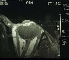
"Movement of water in the anterior chamber of the rabbit eye in response to topical agents for glaucoma therapy was investigated using high-resolution T2-weighted surface-coil magnetic resonance imaging (MRI) aided by a T2 contrast agent, oxygen-17 water. This water-tracer was applied topically which entered the anterior chamber within 10 min. The kinetics of its dissipation from the anterior chamber was recorded with serial T2-weighted MRI. The data represented the total water flow rate in the anterior chamber.

"We tested the effects on water flow of an alpha-adrenergic (phenylephrine), a miotic (pilocarpine), and a beta-blocker (timolol). The results showed that phenylephrine reduced more than 2/3 the normal water flow rate, pilocarpine accelerated the initial flow rate ca. 2-fold although oxygen-17 water did not fully dissipate, while timolol had little or no effect. These results suggest that water flow in the anterior chamber is in large a function of the status of the iris surface area and/or iris circulation. "

It should be noted, the flow rate using the water tracer, oxygen-17 water (MW=17.00) is 10 times that with fluorescein. In the eye, water is expected to interact with the transport systems of all tissues, not just the flow indexed by fluorescein. Indeed, we need to have a more comprehensive view of water flow in the eye.

Think "Die Moldau".

5.1 Franz Schubert Symphony No 8

Yes, the Unfinished, I. Allegro moderato in B minor and II. Andante con moto in E major, and then, "That's all, folks."

Eye research will always remain unfinished, albeit in a much different sense. Schubert is long gone (1797-1828); luckily, there will always be dedicated people who try very hard to understand the eye and figure out the cure for eye diseases. So whatever is unfinished is actually an opportunity and a challenge for the newcomers.

In the US, the ongoing areas of research are defined and sponsored by the National Eye Institute/NIH, including retina, cornea, lens and cataracts, glaucoma, strabismus (plus myopia), and low vision. Each of the areas is also supported by non-profit private organizations and the instrumentation/pharmaceutical industries.

A very good example of the translation of NEI-supported research into treatment success is the anti-VGEF therapy. Because of the demonstrated efficacy in the treatment of neovascular AMD, clinical trials have now extended into other neovascular eye diseases, e.g., RAVE (rubeosis anti-VGEF trial for ischemic central retinal vein occlusion) and VISION (VGEF inhibition study in ocular neovascularization). Others are being examined as well that include diabetic macular edema, cystoid macular edema, and proliferative diabetic retinopathy.

On the other hand, we have noticed a gap between neurophysiology and impaired vision. A very versatile yet under-utilized imaging tool has been available since 1993, i.e., fMRI.

Functional magnetic resonance imaging (fMRI) permits visualization and quantitation of blood flow, volume, and oxygenation in the brain during sensory and motor stimulation. There are two variations of this technique, one detects the susceptibility effect from intravenously administered contrast agents, e.g., GdDTPA, and the other oxygen-induced susceptibility and changes in tissue diffusion but without the use of contrast agents. The non-contrast method is preferred because of its total non-invasiveness. Its applicability to brain function research, especially the photically stimulated primary visual cortex has been verified with a number of studies. Indeed, fMRI has been designed to test various visual stimuli, the studies also pave the groundwork for testing higher functions such as color vision, eye tracking and movement, retinal rivalry, suppression, and stereopsis.

An example is correlating cerebral activation with eye blinking.
Notice the upper leftmost image is activation of the orbitofrontal lobe during normal blinking of once per 4 sec. The rest of the cerebral cortex remains quiet until when the blinking is inhibited, then even the visual cortex is activated.

Much more on ocular malfunctions and the brain's plasticity can be learned by using fMRI. For example, correlating fMRI results and retinal lesions can be done with point-to-point projection between fMRI and the patient's visual field defects from, e.g., open-angle glaucoma, hemifield defects, optic neuritis/atrophy, diabetic retinopathy, and AMD. Indeed, it is not even known, for example, how the primary visual cortex responds to central scotoma, let alone the understanding of brain plasticity in association with preferred retinal loci.

What about lazy eye, treatment (L-DOPA?), plasticity...

Indeed, this field is still wide open.

4.11 Broken windshields

Occasionally there are intriguing cases of blurred vision. And the reasons for which become obvious, once the optics is understood. It is all a matter of the broken windshield, i.e., the cornea, really.

Remember that the projection of images onto the retina requires that the cornea, the crystalline lens, and the vitreous all remain optically clear. Any opacities can cast shadows on the retina. The most obvious and commonly seen are cases of cataracts and floaters. Less obvious is corneal anomalies.

Here is one case with a chief complaint of glare. The cornea has previously undergone RK, i.e., radial keratotomy, with 8 incisions on the cornea:
Here you can see 4 bright radiating lines. They are part of the 8 RK incisions. The cornea is thinner under each cut thereby allowing more light to pass through. This is the reason why this patient sees star burst when looking at any point source of light, e.g., headlights from the approaching automobiles. The edges of the upper left incision is also irregular and it passes over the macula - not an agreeable situation for driving at night.

Yet another example is the multiple dark shadows seen in the image below:
And the source of these dark spots is shown in the following image:
The white areas are a form of corneal dystrophy. The opacities may decrease or increase in intensity causing vision change.

Other types of not-so-subtle broken windshields include:
This is a case of recurrent corneal erosion with pain and decreased vision. And in another, a very common case of superficial punctate keratitis (SPK):
The tiny green spots are areas with epithelial defects, often appearing in very dry eyes.

And of course there are infiltrates from inflammation due to contact lens over-wear:
In this case, an infiltrate slightly to the left of the pupillary area is detected.

Most superficial corneal defects from infection and inflammation can be treated medically and the recovery is almost all total.

Irregular corneas that severely limit vision from, e.g., keratoconus or corneal scarring, may need to be replaced with donor corneas.

Wednesday, December 12, 2007

4.10 Itchy eyes

Allergic conjunctivitis is quite common. It can be seasonal or perennial. It is a Type I hypersensitivity, an inflammation of the conjunctiva resulting from an immune response to allergens.

The main symptom of allergic conjunctivitis is severe itch. The eyes also look red and irritated. Very simply, the allergens (from cat dander, house mites, pollens, or chemicals, etc) sensitize the T- and B-cells to produce IgE antibodies. IgE then binds and activates the mast cells. The allergens then further cause the rupture of activated mast cells, thereby releasing histamine and other chemicals.

The itchiness is a response of nerve endings to histamine which also causes vasodilation and blood vessel leakage - hence the redness of the eyes and the formation of conjunctival follicles. So the general principle of treating allergic conjunctivitis is to avoid exposure. Failing that, to use a topical fast-acting antihistamine or a slower-acting mast cell stablizer, or even better, a combination antihistamine-mast cell stablizer. And in severe cases, to treat first with topical anti-inflammatories followed by the anti-allergy drops.

The most commonly prescribed combination eyedrops include Patanol, Zaditor, and Optivar. The newer mast cell stablizers include Alamast and Alocril. Sometimes low concentration soft steroids such as Alrex can also be used on a long-term basis. We, however, favor the combination drugs for their efficacy and safety.

4.9 Missing visual fields

Occasionally, a patient comes in complaining of severe headaches and, at the same time, part of his/her visual field is missing. A quick trip to the ER via ambulance is a good idea. In less acute cases, it is actually possible to figure out what the problem maybe - by plotting the visual fields.

It is expected that some parts of the visual fields are lost from retinal diseases such as POAG , RD and diabetic retinopathy. However, many other losses are behind the eyeball, from impairments to the visual pathway. An it can be anywhere from the optic nerve, to the optic track, the lateral geniculate nucleus, the optic radiation, and the visual cortex. Any problem along the way exhibits a unique pattern of the field loss.

In all textbooks, the lesions (indicated by a - g in the diagram below) and their corresponding field losses are depicted in a schematic such as the following:
(www.unmc.edu/Physiology/Mann/mann7.html)
And a more anatomically correct representation, looking at the base of the brain, is shown below:
(From nmr.mgh.harvard.edu)
By examining the visual fields, the location of the lesions becomes apparent. The real question is then what the nature of the lesion is. Most times it is either a tumor or some cerebrovascular diseases. An urgent referral to a capable neurologist is therefore in order.

Tuesday, December 11, 2007

4.8 The "flying mosquito" syndrome

Floaters, known as the "flying mosquito syndrome" in Chinese, are little specks, threads, or tiny cobwebs that you see against a blank wall or while looking at the sky. And they move with your eyes. There are many different kinds of floaters, all of which deserve close scrutiny.

First, a quick review of the structure of the vitreous. The vitreous occupies the bulk (80%) of the eyeball. It is 99% water and has a lattice of collagen fibers with coils of hyaluronic acid distributing within the structure. Essentially, the vitreous is an optically clear gel. In the aging eye, parts of the vitreous become liquefied, and the collagen fibrils condense to form the floaters.

An example of this type of floaters is shown below:
Notice the tadpole-shaped shadow in the 2 o'clock position. Cataracts also cast small shadows which are more centrally located, i.e., in the macular area. Floaters are more noticeable if they are situated close to the retina. However, not all intra-vitreous opacities are perceived by the patients. A good example is the tiny soap-like particles known as the asteroid hyalosis:
These particles usually appear in only one eye. Sometimes it is an indication of (often undiagnosed) diabetes, so a blood glucose test is recommended.

Sudden onset of floaters can be a sign of trouble. A good example is tractional retina detachment (RD) during which, a blood vessel is ripped open. The blood cells appear as a sudden shower of floaters when they enter the vitreous. Retinal detachment requires surgical repair, that in itself is an entire branch of ophthalmology. An image of RD is shown below:
(This one image courtesy of Optos)
And the encircling scleral buckle that helps the retina to re-attach itself is seen as a black circular band in this post-op RD:

Again, the image may look like a total mess, the central vision is actually preserved.

Then we have vitreous hemorrhage from a leaking artery:
Notice the origin of the hemorrhage (the dark red spot) is next to the optic disc in the 5 o'clock position. This hemorrhage actually extends into the vitreous seen by the patient as a large floater. A stated before, extensive vitreous hemorrhage requires vitrectomy in order to regain vision.

Finally, a very common condition known as posterior vitreous detachment (PVD). PVD affects 50% of people over 50. Typically the patient sees a sudden appearance of flashes together with a long curvilinear or a circular floater. Sometimes PVD is accompanied by hemorrhage and/or RD, but mostly quite benign except for the annoying floater. One such is shown below:
This circular floater (which appears oval, a sideway view) is initially part of the vitreous attachment to the optic disc, in other words, a rim, which is pulled off during the vitreous separation from the retina. Luckily, no blood vessels are disrupted this time. Once the traction is relieved, the flashes are no longer seen.

So sudden onset of floaters must be treated as an emergency and the etiology identified.

4.7 I see double

Sometimes, a patient self-refers because: "I see double". Double vision is quite disturbing and debilitating at the same time.

Diplopia is usually sudden-onset, and is a manifestation of numerous possible major systemic diseases. It is to be distinguished from squint in children (Section 3.3.2). We will now examine this issue closely. First, the control of the eye movement:
(from health.howstuffworks.com)
The above diagram shows the six extraocular muscles (EOMs) of a left eye. You can imagine a mirror image as the right eye. The muscles must work in coordination to maintain binocular vision, i.e., fixation at the same spot.

Let us take a simple situation, say, you are looking to the left. Then for the left eye, the lateral rectus contracts, while the medial rectus relaxes. At the same time, for the right eye, the medial rectus contracts and the lateral rectus relaxes. At all times, the two eyes are locked in for looking at the same point/object. If you now see double, apparently this coordination is broken. And the underlying reason(s) must be examined both neurologically and neuroradiologically.

Other than a direct trauma to the muscles or a result of eye surgery that is readily apparent from history, one must start from: which muscle is affected. And deduce from it, which cranial nerve is involved. Basically, the 4th nerve innervates the superior oblique, the 6th innervates the lateral rectus, and the rest by the third nerve. Again, take the lateral gaze example, if there is a 6th nerve palsy affecting one of the two lateral recti, then the eyes will not be able to look to the left or the right without seeing double. The question is then what happened to the 6th nerve on its way from its origin in the pons of the brain stem to the orbit and the lateral recti. You'll need to entertain the possibility of multiple sclerosis, mysathenia gravis, diabetes, circulation problems, hypertension, tumors, and other known and unknown causes. Indeed, any of these can disrupt the course of the 6th cranial nerve. Sometimes, the etiology remains unknown. And often the diplopia resolves without any intervention in a few months, especially if the primary cause is diabetes or hypertension.

[Note: Hypertension, with the increasing intracranial pressure affects more frequently the 6th nerve; whereas diabetic infarct, more the 3rd nerve. Diplopia, then, is a sign of poor control of blood pressure and blood glucose, respectively.]

And obviously the same workup goes for 3rd and 4th nerve palsies, except the former is usually accompanied by drooping eyelids (ptosis) and eye/head pain, while the latter is usually benign.

Similar to vertical squint/strabismus, often people with sudden 4th nerve palsy eventually learn to tilt their heads to avoid diplopia. The temporary relief of double vision can be a simple patch for one eye or a large amount of prism in the spectacles.

It is of course the eye doctor's job to perform a thorough evaluation. The patient is then referred for further consult for the subsequent management by appropriate specialists.

4.6 Huge pupils

The size of the pupil does change with age, from somewhat small in infants to 3-5 mm in adults, then it becomes smaller again in older people. It is a change in the muscle tone of the iris sphincter and dilator. In a dilated eye exam, eyedrops are used to paralyze the sphincter and stimulate the dilator to maximize pupil size.

Sometimes we see unusually large pupils in children. These are cause by medication, i.e., medicine-induced mydriasis (about 8mm pupil size). Seen in children treated for ADHD, for example.

According to a 2003 CDC report, 4.4 million kids between 4-17 years old have been diagnosed with ADHD, and 2.5 million of them received medical treatment. The commonly prescribed medications, Adderall (an amphetamine) and Strattera (a norepinephrine re-uptake inhibitor), all have a long list of side-effects. And among them, mydriasis - from a sympathomimetic effect causing contraction of the iris dilator muscle. As a result, the angle narrows which may cause or acerbate narrow-angle glaucoma. In severe cases, the patients suffer from debilitating headaches and photophobia.

2.5 million is not a small number, that is probably why some of these kids show up for eye exams because of blurred vision and reading difficulties due to decreased accommodation. So the scholastic performance may suffer albeit with improving behavior. A paradox of sorts. Unfortunately, there is currently no effective management for this problem.

As they say: more studies are needed.

4.5 Sleeping with contacts

Contact lens over-wear is a problem of non-compliance. While some lenses are designed for overnight wear, most are not. Even the ones approved for 30-day continuous wear have some limitations as well.

In most cases, the patients report being too tired to take off the contacts the previous night. And this morning the eyes are in pain. Often the patients wait a few days hoping the eyes self-heal before seeking care. The root cause is still oxygen deprivation (or hypoxia) made worse during sleep when the eyes must close.

In mild cases, there is inflammation of the cornea, and in severe cases, additional microbial infection that may lead up to corneal ulceration. Treatment is case-dependent from palliative to anti-inflammatory and/or antibiotic therapy.

Now an opportunity to discuss contact lens compliance. It is often forgotten that contact lenses are a medical device. As such it is subject to the rules and regulation of the FDA. The doctor will always does his or her best to fit you with lenses that allow good vision and good comfort, within the prescribed wear-time. And it is the patient's responsibility to adhere to the wear schedule and keep the lenses clean.

Here is a quick list:

1. Replace contacts as required;
2. Replace or disinfect contact lens storage cases periodically and never allow solution to dry up in the cases;
3. Wash your hands with soap and water and rinse thoroughly before handling the lenses;
4. Do rub the lenses both sides, each side at least 10 sec even if you are using a "no-rub" multi-purpose solution and make sure the bottle is capped after each use;
5. True no-rub solution is H2O2-based which performs better than others in terms of disinfection;
6. Stop wearing lenses if the eyes look red and/or if you feel any discomfort;
7. Seek medical help as soon as possible, do not self-medicate.

And whatever you do, don't over-wear your contacts!!

Monday, December 10, 2007

4.4 Dry eye

The following is played over and over in many offices around the world:

Doctor, at the slit-lamp: "Mrs Smith, the reason why your eyes are sore is because you have very dry eyes."
Mrs Smith, moves back, in disbelief: "How can that be, I tear very easily, all the time, too..."

They are both right, of course. The doctor is summarizing what he/she gleaned from symptoms and signs. The patient, on the other hand, is describing reflex-tearing caused by irritation from dry eyes.

The tear fluid is actually composed of more than just water. It has three layers, a very thin top layer of lipids, a much thicker layer of water with all sorts of solutes, and a thinner layer of mucus that interacts directly with the corneal epithelium.

Imagine a very shallow lake, with in- and out-flows, sitting on top of your cornea. If the water source slows to a trickle, then the lake slowly dries up. You can partially plug up the drain and maintain a certain water level. This is an over-simplification of course; although in clinical practice, the plugs are known as punctal plugs. And indeed if the lacrimal gland is inflammed/damaged, then the production of tears is reduced.

When the eye is chronically dry, the surface structures tend to weaken. Often you'll see subconjunctival hemorrhage. A patch of bright red spot next to the cornea (see iamge below).
In other words, the blood vessels have become so fragile, and with a little stress, e.g., contact lens insertion, sneezing, strained bowel movement, one of the blood vessels will break open and the blood leaks into the adjacent conjunctival tissue. It usually resolves on its own in a week or so. Of course there are other underlying causes for this type of hemorrhage such as blood diseases, blood-thinning medication, etc. Differentiation is needed.

So what causes dry eye? Other than a reduction in tear production, if the top lipid layer becomes so thin as to be broken, then water evaporates very quickly. This happens when there is a malfunction in lipid-generation from glands such as the Meibomian glands. This is part of the inflammation of the lid margin (aka blepharitis). This condition is further aggravated if you work in a dry environment and stare at the computer all day long.

Race seems a big factor as well as most Asians suffer from dry eye. Their tear break-up time (around 4-5 sec) is much shorter than that of the Caucasians (around 20 sec).

Some medicine such as birth control pills, levoxyl, high blood pressure pills etc can also cause dry eyes. The major ones are from (1) contact lens wear and (2) Sjogren's syndrome. Contact lens disrupts the integrity of the tear film. Sjogren's is an auto-immune disease (confirmation through blood tests for ANA and rhrumatoid factor), seen mostly in menopausal women, with dry eyes, dry mouth, and often arthritis.

The diagnosis of dry eye can be simply from patient history, supplemented with fluorescein and/or lissamine green staining - for signs of epithelial defects. In severe cases, measurements of tear break-up time, tear film integrity, and tear production rates are also done.

The treatment of dry eye has made quite a bit of progress, especially the type that responds to anti-inflammatory therapy. In this type, a pulse dosing of topical steroid is tested first. If effective, then a long-term treatment with topical soft steroids, oral omega-3 fatty acids, or cyclosporine eyedrops should follow. And when needed, artificial tears are also used.

Sunday, December 9, 2007

4.3 Diabetic retina

Diabetes is a nasty disease of ischemia. There is no cure for it just yet. It comes in two forms, the familiar Type 1 and Type 2. There used to be an aging factor for Type 2 (i.e., adult-onset), now even children develop Type 2 diabetes. Long-term diabetes leads to many complications in the eye including diabetic retinopathy, cataracts (posted previously, see Section 2.2.3), and sometimes, neovascular glaucoma.

Let us ponder this wide-field retinal image first:

In the center, there is an island. Which is surrounded by a sea of bright spots. In the middle of the island, you can see the macula, to its left is the optic disc, and to its right, a suspiciously looking dark-red area. Also within each bright spot, there are black specks. Yes, this is a diabetic retina after a pan-retinal laser photo-coagulation (PLP) treatment. The bright spots are laser burns and within each spot, pigments from pigment epithelium. The purpose is to stop bleeding as that in the dark-red area. Usually there are 1,000 burns.

Why is this treatment necessary? Well, we need to backtrack a little. Because of the structural changes of the blood vessels in a diabetic retina (in the whole body, in fact), we can see micro-aneurysms (often known as background or non-proliferative diabetic retinopathy). Then three things happen: (1) the delivery of oxygen is inadequate; (2) the blood vessels sometimes leak; and (3) the macula may become edematous. These get progressively worse as time goes on. Eventually, the ischemia gets so bad, new blood vessels proliferate to provide more oxygen. So now we have the proliferative diabetic retinopathy. These abnormal vessels often break and blood will leak into the vitreous necessitating a vitrectomy (surgical removal of the vitreous). PLP is therefore performed to stop these hemorrhages. The image above may look terrible, yet the patient still retains central vision, even though peripheral vision is somewhat compromised. This is still far better than a total vision loss.

To avoid or delay these complications, tight blood sugar control is absolutely crucial. So if you are a diabetic, do yourself a big favor: Follow your diabetes doctor's instructions religiously. And pay attention to your Hb A1c level and monitor your own blood glucose closely.

4.2 So you have high IOP

First, some pictures of the optic disc:

(POAG with large cupping occupying 90% of the disc area)

(Normal optic disc with no cupping)

Glaucoma in adults actually has many different types. It most commonly refers to POAG, primary open-angle glaucoma, which can be familial. In Asia, we often see acute angle-closure glaucoma in a hospital ER. It is a true medical emergency, which also can be a consequence of chronic narrow-angle glaucoma. Then we have secondary glaucomas from neovascularization (e.g., rubeosis in diabetes), pigmentary dispersion, phacolysis, pseudoexfoliation, uveitis, or trauma. And of course, there is this somewhat mysterious normal-tension glaucoma (NTG) in which, as the name indicates, the intraocular pressure (IOP) is actually within the normal range and the angle is open as well. Interestingly, NTG seems to be more common among the Japanese and it is often associated with migraines and cold hands/feet.

This "angle" refers to the junction between the cornea and the root of the iris where the intraocular fluid drains, into the trabecular meshwork and then into Schlemm's Canal and the venous system.

Acute angle-closure is a condition where an adhesion between the back of the iris and the front of the crystalline lens causes a forward bulging of the iris proper (see picture below). Chronic narrow-angle is similar but without the iris-lens interaction. In both cases, the angle closes or narrows. The fluid can no longer drain properly, resulting in an elevated IOP. The solution is, in the acute case, to reduce the IOP pronto, followed by laser iridotomy - after the pressure is under control. And in the chronic case, medical control of the pressure as in POAG or laser treatment. The purpose of laser iridotomy is to open a hole in the iris proper to allow a direct fluid flow from the posterior aqueous chamber into the angle. It can be and probably should be done on a prophylactic basis.

(Modified from najafimd.com)
Here, we'll touch upon the most recent advances in diagnosis and treatment of POAG. Indeed the evaluation of POAG is now heavily technology-driven and the wide availability of efficacious anti-glaucoma agents is astounding.

First, the diagnosis. In the not so distant past, IOP is used as the primary if not the sole index of POAG. To this day, patients still request a "glaucoma test" which is essentially a pressure check using a gauge (i.e., a tonometer). Now it is known that a high IOP does not always lead to POAG and the pressure readings must be corrected based on corneal thickness. For example, patients after LASIK thinning of the cornea will have a falsely low IOP. And the same applies to people with thin corneas. The corrected IOP, if high, must still be complemented with an evaluation of the optic disc for evidence of "cupping" (see pictures at top of page) and nerve fiber defects, followed by a visual field test for any losses. A definitive diagnosis of POAG (and NTG) must be based on all three. The "cupping" is a depression or excavation of the optic nerve head caused by elevated IOP, which also damages the retina and the optic nerve leading to the visual field loss (see below: from A to D, progressive loss of visual fields indicated in black).
(from www.aafp.org)
The corneal thickness is measure with a pachymeter. The angle and the optic disc can both be imaged with OCT (Optic Coherence Tomography). And the visual fields plotted with perimeters of various designs. The treatment of POAG and NTG is fairly straightforward. Anti-glaucoma drugs now include the following:

Prostaglandins: We now have Xalatan and Travatan (both esters) and Lumigan, an amide. One drop a day can reduce the IOP by 30%.

Beta-Blockers: There are a few of them, Betagan, Betoptic, Timoptic, Betimol, and Istalol. It is well-known that beta-blocked must not be used on patients with respiratory illnesses, such as asthma, or cardiac issues such as congestive heart failure - or risk death.

Alpha-adrenergic agonists: Alphagan is an example - to be used as a second line drug if the first line drugs, prostaglandins and timolol, are not tolerated by the patient .

Carbonic anhydrase inhibitors (CAIs): CAIs can only reduce the IOP by 15%, so a daily dosing of 2 to 3 times maybe needed. We now have Azopt and Trusopt that are often used together with timolol.

Indeed, proper diagnosis, optimal dosing, patient compliance, and vigilant follow-ups can prevent vision loss in almost all cases of POAG and NTG.

Of course, there are cases of recalcitrant glaucoma, especially that of neovascular origin (as a complication of diabetes or hypertension), which cannot be controlled by medicine. Surgical intervention as that for congenital glaucoma is then performed. The outcome is somewhat guarded, however.