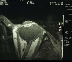
Occasionally, a patient comes in complaining of severe headaches and, at the same time, part of his/her visual field is missing. A quick trip to the ER via ambulance is a good idea. In less acute cases, it is actually possible to figure out what the problem maybe - by plotting the visual fields.
It is expected that some parts of the visual fields are lost from retinal diseases such as POAG , RD and diabetic retinopathy. However, many other losses are behind the eyeball, from impairments to the visual pathway. An it can be anywhere from the optic nerve, to the optic track, the lateral geniculate nucleus, the optic radiation, and the visual cortex. Any problem along the way exhibits a unique pattern of the field loss.
In all textbooks, the lesions (indicated by a - g in the diagram below) and their corresponding field losses are depicted in a schematic such as the following:
And a more anatomically correct representation, looking at the base of the brain, is shown below:
By examining the visual fields, the location of the lesions becomes apparent. The real question is then what the nature of the lesion is. Most times it is either a tumor or some cerebrovascular diseases. An urgent referral to a capable neurologist is therefore in order.
Subscribe to:
Post Comments (Atom)


No comments:
Post a Comment