 One wonders if both Felix Purcell (1912-1997) and Edward Bloch (1905-1983) had experimented with magnets (see above) when they were little. They did, however, share the Nobel Prize in Physics in 1952 for independently creating experiments for the observation of NMR (nuclear magnetic resonance).
The growth of NMR research especially in the past two decades is nothing short of spectacular. It is a field driven entirely by the magnetic field strength (B0) which determines the operational frequency or Lamor frequency (ω0
ω0
γ is of course the immutable gyromagnetic ratio (or magnetogyro ratio - depending on whether you are a physicist or a chemist) unique to each NMR nucleus. The table below is a quick glance of the Larmor Frequency at a field strength of 1 Tesla (last column on the right):| Particle | Spin | wLarmor/B s-1T-1 | |
| Electron | |||
| Proton | |||
| Deuteron | |||
| Neutron | |||
(Note: the study of electron spin is ESR - Electron Spin Resonance or now known as EPR - Electron Paramagnetic Resonance - an entirely different ballgame from NMR)
In the old days, field strength was represented by proton frequency. For example: what was a 400MHz (or megacycle) magnet is now known as a (400/42.5781=)9.4 Tesla magnet. Because of the notoriously low sensitivity of NMR nuclei, much improvement in signal generation relies on increasing the magnetic field strength. The higher the field strength, the more the signal intensity (or higher signal-to-noise ratio). For example, for proton, an increase from 300 to 400MHz yields an improvement in S/N of (400/300)2 = 1.78 fold. Building ultra-high-field magnets is a competitive sport for the manufacturers of course.
What are these NMR nuclei? Well, any nucleus with an odd number of protons or neutrons (or combination) qualifies; for example, biologically relevant nuclei include 13C, 31P, 1H (proton), 23Na, 17O, 15N, 19F, and deuterium (a nucleus with spin number I=1). Of these, 31P and 23Na are both 100%, and 1H 99.98% naturally abundant. The others are only a fraction of around 1% or less and are usually used as labels; for example, 13C can be labeled at either C-1 or C-6 position of glucose and the glycolytic products can thus be traced. On the other hand, proton is part of the ubiquitous water molecule, and 31P that of ATP and other organophosphates.
All these nuclei when placed in a magnetic field will precess at certain ω0: depending on the γ of each nucleus, as mentioned above:
If a radiofrequency (rf) perpendicular to the north-south direction, or conventionally, the z-axis of the magnetic field is applied at the frequency of a nucleus of interest, then this nucleus will be tipped into the x-y plane. When the rf is turned off, the nucleus will begin to return to its original ground state through a process, known as relaxation. Relaxation is defined by longitudinal relaxation time (T1) and transverse relaxation time (T2). And the signals produced in the rf field are known as the FIDs (free induction decays) which can be picked up by an antenna and processed (through Fourier Transform from time into frequency domain) to generate NMR spectra.
The above is a typical high-field vertical-bore superconducting magnet used in NMR spectroscopy. It requires periodic feeding of liquid nitrogen and liquid helium. A probe is inserted from the bottom and the sample (in an NMR tube) loaded from the top, controlled by compressed air. A computer console is situated nearby for shimming, pulse sequence delivery, signal collection, spectral generation and printing.
An NMR probe is the heart of the spectrometer, it fits vertically into the bore of the magnet. They come in different bore sizes as well, e.g., 2, 5, 10, and 20mm diameters are common. These probes can be broadband or single frequency.
 Before insertion, it needs to be optimized by using tuning and matching rods with the aid of an oscilloscope. Tuning is simply to make sure the probe is set to the frequency of interest. And matching is to ensure the impedance is equal to the external electronic circuitry so that maximal power can be transferred. These two operations are actually inter-related:
 Shimming is an art, its purpose is to maximize the magnetic field homogeneity which greatly affects spectral resolution. Some "NMR jocks" are known to take extraordinarily long time (e.g., forever) to shim the magnet - to perfection. And a lot of them know how to read the FIDs even before FT. The resulting spectra are often strikingly well-resolved. NMR jocks are a special breed. Much like computer hackers, NMR jocks also survive on pizza and coke, except the food and drink are paid for with cash, as their credit cards are usually erased by the magnets. (Try and explain this to the pizzeria owners.)
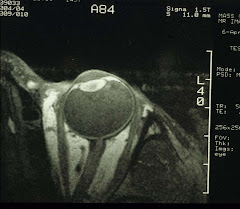
Next, we will examine a few examples of NMR spectroscopy of ocular tissues - before entering the field of MRI (magnetic resonance imaging).


