Friday, January 11, 2008

7.6 T. rex vision


Have you ever wondered how the dinosaurs saw when you look at their immense fossil heads with huge empty eye sockets?

Of course, all the soft ocular tissues are long gone, so it is impossible to know the gross anatomy of the eye, let alone how many kinds or the density of photoreceptors in the retina. Without the information, all studies are essentially best guesses.

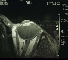
T. rex and its relatives, the theropod dinosaurs, all have a huge head, small fore-limbs with sharp claws, and walked or trotted on powerful hind legs. Based on the position of the orbits, it is possible to estimate the overlapping visual fields of the two eyes. It turns out that two possibilities exist, one with a 20° overlap (similar to that of the crocodiles) and the other 45–60° (similar to that of the birds). T. rex belongs in the latter group with a 55° overlap (see image at top).

So what does this overlap mean? Most likely for stereopsis at close range. According to some paleontologists: if you are a predator as the T. rex, it is a good idea to see what you are biting at. T. rex is known to leave its well-placed tooth marks on its victims. On the other hand, a scavenger needs only to know where the meal lies; and a wider peripheral visual field is advantageous for scanning the horizon, in case some big dangerous looking T. rex is lurking nearby.

The above seem reasonable. The next assumption is big eyes must have excellent vision. In fact, there has been some exercise fitting an enlarged version of reptile or bird eye into the eye socket of a T. rex and project what its vision could be. Some claimed T. rex had 13 times better acuity than humans. From the retinal point of view, this appears unlikely. The eyes of a T. rex maybe several times larger than that of the humans, it is not the number but the density of the of photoreceptors that determines visual acuity. That is, if T. rex did have a fovea as in the humans. Perhaps it had multiple foveas each for a different visual function for all we know (the eagles have two, for example). Without an actual sample of the retina, it is simply not possible to draw any definitive conclusions.

So how did a T. rex see? Very well, thank you very much.

Friday, January 4, 2008

7.5 Flowers, where?

Do animals have color vision or even how they see - no one really knows. We can, however, make some guesses. Dogs have two types of cones, humans three. So dogs don't have the same color vision as humans. Both dogs and humans have two types of photoreceptor cells, i.e., rods, and cones. Birds have three: cones, rods, and a mixed type, so some birds can see more colors (e.g., humming birds), while others can see more light at night (e.g., owls) than humans/dogs. Vision is not created equal at all .

Humans normally perceive color within the visible light spectrum of 380-750 nm. Not everyone does, of course. It is safe to say that one in twenty guys (5%) is color blind or more precisely, red-green deficient. This is caused by an X-linked recessive gene. Females can be R-G deficient, too, though much less common - it is hard to receive two copies of the rare gene. An even less prevalent form is blue-yellow deficiency and the most rare is total color blindness. Most eye doctors have never seen the latter two types in their entire professional careers.

Red-green deficiency is just that: the patients cannot readily differentiate, e.g., pink flowers from green leaves, or red from green apples (see images below). They will have trouble with traffic lights if the lights are arranged in an unfamiliar fashion (e.g., horizontal rather than the usual vertical with red at top). Not to mention the inability to tell whether a severe case of sunburn is coming on, a woman is wearing lipsticks, or a piece of steak is too well-done. And forget about reading litmus test results or color coordinating clothing. Otherwise the visual functions are quite normal.

The remedy, although not ideal, is to wear red-tinted contact lens (known as the X-Chrome lens) in one eye only. The patient can actually identify Ishihara color vision test plates just like any normal-sighted person. If you are R-G deficient, a DIY way is to cut out a piece of red mylar file cover, laminate it onto one of the lenses in your spectacles. Put your glasses on, look around, and be surprised at what you have been missing all your life.

(Top: red and green apples; bottom: as seen by a R-G deficient person [from answers.com])

There have been attempts at gene therapy repairing color blindness; although it is still unclear if any success (or even necessary).

If a normal-sighted person begins to lose color differentiation and who is afflicted with one or more of the diseases listed below. Then that is not so good because it indicates damages to the optic nerve and/or the retina. And the list is relatively long:

Eye diseases include POAG, AMD, and retinitis pigmentosa. Systemic diseases include Alzheimer's disease, diabetes, liver disease, chronic alcoholism, multiple sclerosis, Parkinson's disease, leukemia, and sickle cell anemia.

Many drugs also can have side-effects that involve disturbance to color vision. Usually your prescribing doctor will have informed you so.

And chemical poisoning by carbon monoxide, carbon disulfide, styrene, and lead can cause disturbance to color vision as well. The most famous example is Vincent van Gogh (1853-1890) who loved to paint in intense yellow (you all recall his "14 Sunflowers in a Vase"). Mr van Gogh was probably a walking medicine cabinet. In his time, he was treated for various ailments which have been theorized to be, take your pick, epilepsy, bipolar disorder, chronic sunstroke, acute intermittent porphyria, lead poisoning, and Ménière's disease. From his paintings, poisoning by absinthe liquor, digitalis, or even lead is indeed quite possible.

Wednesday, January 2, 2008

7.4 Tako sashimi

If you are a Jerry Lettvin fan, please skip to paragraph 2. Mere mortals, please read on:
RECIPE
Remove octopus guts, eyes, and beak and freeze for at least 72 hours.
Thaw and steam for 30 minutes or until done.
Cut off the tentacles and with a very sharp knife cut them into 1/8 inch slices at a 45 degree angle. Slice the belly into 1/4 inch strips.
Mix wasabi with soy sauce.
Dip octopus and enjoy.
(For more, visit http://www.freediver.net/freedivelist/recipes.html)
Save the eyes. Or visit your local Japanese restaurant, have some sashimi, and ask for octopus eyes while there. Why? A comparative anatomical study.

It is often mentioned that the octopus eye is structurally similar to the human eye, complete with cornea, iris, lens, vitreous, and retina. Presumably, it has sharp, color, and 3D vision, and is able to differentiate shapes.

However, a close examination shows that its crystalline lens has a fixed focal length like that of a camera, so the octopus focuses by moving the lens close to or away from the retina (some say by changing the shape of the eye globe). Also, the coordination of the octopus eyes is via the statocyst so the slit pupil of each eye is always in the horizontal position. Probably the most interesting feature is the retina: the photoreceptor cells are directed towards the light source.

If you go back to Topic 2.3.1, you'll see the human rods and cones are oriented against the light source, in other words, the photoreceptors are pointing towards the choroid/sclera, while that of the octopus, towards the vitreous. In terms of the efficiency in photon capture, the octopus obviously has the upper hand. Strange, huh? In fact, this has been used as the evidence of a design flaw of the human eye.

Actually, the octopus lives in (sea)water which has a refractive index of 1.33 and the sunlight is refracted and polarized when it enters water (especially at dusk and dawn). The octopus hunts at dusk. To see a prey such as a jellyfish, it will need a crystalline lens with even higher refractive index (than 1.33) in order to focus properly - because the corneal refractive power is neutralized by the water outside and the aqueous humor inside the eye. And equally important, it will need polarization vision (which actually works well with horizontal slit pupils). Polarized light is less intense and spectrally shifted, yet with more contrast, so it allows the octopus to better appreciate the pattern that leads to the capture of, e.g., a well camouflaged crab. Polarization vision will require the photoreceptors to directly analyze the incident light or the visual information is lost. The photoreceptors therefore should be oriented towards the light.

Humans can always wear Polaroid sunglasses, e.g., to see comfortably through light reflected and polarized by the surface of water/ocean. If you are a diver, try diving with your polarizers on at dusk and see if you can spot a Portuguese Man-of-War floating by. Report back after you have recovered from the pain. An octopus on land will need heavy prescription (for high myopia) just to navigate.

The take-home message: All eyes are developed to serve the one and only purpose: to see well to survive, wherever the rest of the body may reside.

Thursday, December 27, 2007

7.3 Aji-no-moto

Aji-no-moto, the "essence of flavor", commonly known as MSG (monosodium glutamate), has been a fixture in kitchens of all sizes, all over Asia, since 1909. A few grains of the chemical turns "clear water into chicken soup" as the advertisement goes. And in fact, no ill physical effects have been noted until 1968, when reports surfaced in the US about physical discomfort suffered by some, after consuming Chinese food. Despite conflicting data from several studies later on, the label of "Chinese Restaurant Syndrome" stuck. To this day, in American culture, any discomfort, including postprandial stupor, from eating out, is automatically attributed to CRS. And restaurants began to offer MSG-free meals since that time, even though the replacement, the soup stock, is quite rich in MSG.

Generally, the symptoms are mild that include transient headaches, flushing and sweating. It is unknown if some patients are ultra-sensitive to even a small dose of MSG; although it does not make sense why any reasonable immune system should attack glutamic acid, which is actually one of the 20+ essential amino acids, the building blocks of protein - unless it is something neurological as some have noticed later (see below). In any case, a normal dose of vitamin B6 seems effective in preventing or staving off CRS.

What's MSG, or more accurately, glutamic acid, got to do with the eye? Let's make a long story suitably short:

Glutamate is actually a major excitatory neurotransmitter in the brain. It is involved in the very complex behavior of NMDA (N-Methyl-D-Aspartate) glutamate receptors. It turns out that excessive glutamate can damage these receptors. In fact, excitotoxicity due to glutamic acid has been proposed to be the cause of neuron destruction in patients with strokes (and other forms of brain ischemia).

Maybe this toxicity can explain the CRS? No, not really, because there is a blood-brain barrier, aji-no-moto cannot enter the brain without invitation/breach. The source of the endogenous glutamate is the injured local cells, causing excessive neuronal excitation of their neighbors, hence the increasing tissue damage. The real culprit of CRS still remains unknown; although it is most likely not dietary MSG.

But wait, since someone has mentioned ischemia and the retina is part of the central nervous system, i.e., the brain, doesn't it make sense that glaucoma/diabetes related damages to the retina also can be blamed on glutamate excitotoxicity? Maybe so. And a lot of research projects are examining all facets of this issue. Already neuro-protection is a topic of major interest in the management of POAG. For example, drugs for treating Parkinson's disease are being used/tested in a number of advanced cases.

On the other hand, it is also very important to realize that as part of the blood-brain barrier, there is the blood-ocular barrier, which prevents the entry of exogenous glutamate (e.g., MSG in Chinese food) into the retina. And even in the lab, an extremely high concentration of glutamate is needed to produce direct cellular damages to the retina. Again, we can rule out dietary MSG in all ischemia-related retinopathies.

Often in the lay press, a few keywords are lumped together to create a seemingly scientific report. In the above case, putting MSG and neurotoxicity together in an uncritically written article can lead the reader to erroneously conclude that "eating out at a Chinese restaurant can damage your brain/retina". On the other hand, without the press coverage and the ensuing publicity, research on glutamate and its excitotoxicity would not have advanced so far. Ultimately, the patients benefit, so the system does work despite its many flaws. Readers must still exercise their own judgment, of course.

Monday, December 24, 2007

Sunday, December 23, 2007

7.2 Harder who?

Next time, when you visit your eye doctor, casually mention that you would like to have your Harderian gland checked. The doctor will look at you quizzically, "Harder who?"

With the long and rich history of human anatomy, you'd think that everything in the human body, big and small, young and old, have all been discovered and carefully documented. Not so. The Harderian gland has escaped attention, even though its existence in other mammals has long been known. In fact, it was first described in 1694 by an obstetrics professor in Basel, Johann Jacob Harder (1656-1711).

Indeed, one time, an excited nuclear medicine research group, who have just developed PET microscopy, informed us a large uptake of a certain compound (we are sworn to secrecy as to its identity) by the posterior portion of the eye, in a living rat. And they have traced it to the Gland of Harder. This uptake was later confirmed and quantified with another study. The excitement, however, was somewhat dampened when told that there was no such gland in the humans, at least not the ones we saw. In our collective knowledge, only one person recalled reading about this gland in non-primate mammals - in the comparative anatomy chapter of Wolfe's Anatomy of the Eye. This gland is hiding posterior to and underneath the eye ball, blended into the fatty tissues of the orbit. Easily missed if you are not looking for it intentionally.

What a shame - as PET (Positron Emission Tomography) microscopy is nothing to sneeze at. PET scans usually show low-resolution whole-body/brain images, to see images of a 3-mm rat eye is no small feat.

In 2006, two biologists in Pennsylvania finally published a paper: "Primate Harderian gland: Does it really exist?" in Annals of Anatomy - Anatomischer Anzeiger Volume 188, Issue 4, 3 July 2006, Pages 319-327. The finding? Indeed the Harderian gland is found in fetal and neonatal stages in humans but is largely absent from adults. And of course, its role is still unknown. (In contrast, numerous functions have been proposed for the Harderian gland in non-primates.)

Who knows why the humans do not have a functioning Gland of Harder, but then we don't have the nictating membrane, either.

Sometimes we run into an unknown. And to have found the answer is like discovering some new species in the deep jungles of Indonesia. Sometimes, however, the findings are to be filed away perhaps for another day. All part of the fun and game of research.