Tuesday, March 25, 2008

7.30 Poetic review

It has been stated: "Science is for those who learn; poetry, for those who know." ~Joseph Roux (1725-1793), Meditations of a Parish Priest

Such a great topic for fMRI study of the brain, too. Perhaps when they think, scientists and poets activate different areas of their brains, and the areas do not usually overlap. If true, then Galileo would not have been able to do the following:

"If Galileo had said in verse that the world moved, the inquisition might have let him alone." ~Thomas Hardy OM (1840-1928)

Often the progress in science results in unintended cultural consequences. A good example is cited here:
The above is a poem celebrating the mid-autumn moon. It was written by Su Shi (蘇軾,1037-1101) invoking the Chinese folklore of Chang-Er (嫦蛾) who flew to and resided on the moon - after eating a Heavenly Peach. In 1969, Neil Armstrong's a "giant leap for mankind" moon-landing spoiled it for all, forever.
This poem still retains its popularity; however, now only the last stanza makes sense, i.e., the moon is seen/shared by all even if they are separated by a vast distance.

The eye in fact figures prominently in numerous poems. Let's see if science has changed anything. We'll now try some American poems:

"Thy fingers make early flowers" by e. e. cummings (1894-1962):
thy whitest feet crisply are straying.
Always
thy moist eyes are at kisses playing,
whose strangeness much
says; singing
(though love be a day)
for which girl art thou flowers bringing?

Moist eyes, huh? See "4.4 Dry eye" and "4.10 Itchy eyes" to understand why some eyes are overly "moist".

"Dust in the Eyes" by Robert Frost (1874-1963):
If, as they say, some dust thrown in my eyes
Will keep my talk from getting overwise,
I'm not the one for putting off the proof.
Let it be overwhelming, off a roof
And round a corner, blizzard snow for dust,
And blind me to a standstill if it must.

The immediate physiological response to dust in the eyes is profuse tearing. This condition often requires flushing the eyes with water or normal saline. Prophylactic antibiotic/anti-inflammatory eyedrops may be needed as well. And if the dust happens to be alkaline in nature, e.g., lime, a quick referral to an eye doctor is not an overwise next move. (On the other hand, blinding snow is obviously harmless, unless you are driving on I-93 North.) For more, see "7.17 Wink, wink".

Some poems about death and the eye are downright spooky:

"I’ve seen a dying eye" by Emily Dickinson (1830-1886):
I've seen a dying eye
Run round and round a room
In search of something, as it seemed,
Then cloudier become;
And then, obscure with fog,
And then be soldered down,
Without disclosing what it be,
'T were blessed to have seen.


Corneal edema immediately before death? This is quite unlikely. Fresh fish have clear eyes which become cloudy with prolonged storage on ice - perhaps this is what Dickinson was referring to? See also: "7.1 Cold fish eye".

"The Hollow Men" by T.S. Eliot (1888-1965):
The eyes are not here
There are no eyes here

In this valley of dying stars
In this hollow valley
This broken jaw of our lost kingdoms
In this last of meeting places
We grope together
And avoid speech
Gathered on this beach of the tumid river
Sightless, unless
The eyes reappear
As the perpetual star
Multifoliate rose
Of death’s twilight kingdom
The hope only
Of empty men.


Transplanting the whole eyes? See "7.21 Never say never?" for details. Plus "7.8 Now you see it, now you don't" just to cover all grounds.

What about poems from the East?

"Fallen flowers" by Li Shan Yin (李商隱, 812/3 - 858AD):
高閣客竟去, 小園花亂飛 (My best friend has just left, the flowers are flying/dispersing in the small garden)
參差連曲陌, 迢遞送斜暉 (From the tortuous paths, I now send off the evening sun)
腸斷未忍掃, 眼穿仍欲歸 (Too sad to sweep off the fallen flowers, still hoping to see the friend's return)
芳心向春盡, 所得是沾衣 (My heart is broken as the abrupt end of Spring, only the petals remain on my clothing)

It can be an accommodation/focusing issue to see so intently. See "7.20 Mr Sulu, take us home" for explanation.

"Wan Shi Sa (浣溪沙)" by Wang Guo Wei (王国维, 1877-1927):
山寺微茫背夕曛,鸟飞不到半山昏.上方孤罄定行云. (In the dim dusk, it was hard to make out the temple, for the birds to fly half-way up the hill; the fleeing clouds were anchored in place by a lone prayer drum)
试上高峰窥皓月,偶开
天眼觑红尘,可怜身是眼中人. (I tried to climb up to the mountaintop to peek at the luminous moon, in a blink of the heaven's eye, I could see the world, and realized sadly that I was also part of this world.)

Heaven's eye? The Hubble telescope? Maybe this:
For less powerful telescopes, see "7.27 Thousand mile eye".

Clearly, the word "eye" sets off a reaction of clinical interpretation that overtakes the entire poetry. Essentially, the one-tracked mind at work. Although, we suppose Carl Sandburg (1878-1967) was being honest who said, "I've written some poetry I don't understand myself."

More to the point, though, is this:
"You can't write poetry on the computer." ~Quentin Tarantino (1963-present)
(Much less translate.)

In any case, we now call for an fMRI brain study. Poets are invited.

Saturday, March 22, 2008

7.29 HAL 9000

Of course you all recognize this scene from "2001: A Space Odyssey" with Frank Poole (l) and David Bowman both hiding in a pod, discussing HAL 9000's increasingly erratic behavior. Unfortunately, HAL was programmed to read lips and "he" was therefore able to learn the astronauts' intention of pulling his plug, and took steps to eliminate both humans. You can see the cyclopean HAL outside the oval window, and a close-up of HAL's eye:
Notice it does not has a clearly defined pupil, unnecessary in a controlled environment such as the interior of a spaceship, we suppose. In George Lucas's Star Wars, C3PO does not even have pupils only vertical slits.
On the other hand, the eyes of the Terminator (by James Cameron et al, 1984) are quite interesting:
A dying Terminator was actually shown with pupil reflexes gradually dimming with the ebbing (machine) life (when it was being crushed by a hydraulic press). It is quite obvious that careful thoughts have been put into the movie plot. Finally, in "I, Robot (2004, based on Issac Asimov's original work)", human-like eyes appeared:
The ultimate is of course an android named Commander Data of Star Trek TNG; although "he" is simply too human to be credible. It was probably too costly to construct a life-like robotic prop for the TV series.

In real-life, machine vision (MV) using artificial intelligence (AI) techniques, such as expert systems, fuzzy logic, inductive learning, neural networks, genetic algorithms, and swarm intelligence, is no longer a nascent field. For automation, MV has applications in manufacturing of semiconductors, electronics, pharmaceuticals, medical devices, automotive and consumer goods, and also in packaging and facial recognition.

The next challenge is the design and construction of autonomous robots (as opposed to "robots" remotely controlled by humans). In fact, true robots are beginning to make its presence felt. It is, however, a bit disconcerting to see Asimo (by Honda) possessing no discernible eye structures at all:
What gives, really.

Arthur C Clarke (1917-2008), the sci-fi visionary, was actually right - HAL-like lip-reading machines are now being developed. They will be capable of translating audio speech into visual speech, based on light reflected from the moving parts of human speech and the reflection collected by, naturally, machine vision. And the purpose for these machines? Hmm, a very good question.

Just to add more intrigue: Each letter of HAL (Heuristic Algorithmic Computer) is one alphabet before IBM, and the significance of which is still unclear.

Sunday, March 16, 2008

7.28 Optic ischemia

Did you know that Johann Sebastian Bach (1685-1750; portrait on a stamp, mit one of the Brandenburg Concertos, see above) was blinded by acute angle-closure glaucoma a few months before he died? Also, Messrs Jose Feliciano, Stevie Wonder, and the late Ray Charles all suffered from congenital glaucoma? And former Minnesota Twins baseball all-star, the Hall-of-Famer, Kirby Puckett (1960-2006) was blinded by glaucoma in his right eye?

What kind of damages you ask? Well, the definition of glaucoma especially that of OAG (open-angle glaucoma) keeps changing. It is a sign of progress; although, the answer to "what is glaucoma?" is no longer straightforward. Perhaps in the next few years, we'll know the whole story. For now, we can only work with what we have.

We'll narrow down the discussion a bit to only open-angle glaucoma: OAG has traditionally been regarded as a topical (as opposed to a systemic) issue. In other words, if the intraocular pressure (IOP) is high, then the eye may suffer pressure-induced damages to the retina and the optic nerve, in the form of ischemia. The treatment is then to normalize the pressure using pharmaceuticals. The problem is in some cases, medicine alone seems ineffective, the disease process continues nonetheless. In low-tension glaucoma, the IOP is actually normal, yet the glaucoma-related ocular damages still occur. So, something else apparently is going on, but what is it? Is it associated with systemic circulation? Shall we now institue systemic neuro-protection? And to complicate the situation even further: It also should be noted that a high IOP does not always lead to OAG.

Even more basic: How do we know optic ischemia is involved? Or more accurately, how do we measure the oxygen level in the optic nerve under high IOP? Short of inserting an oxygen-sensing electrode into the optic nerve itself, there isn't really any good way. One can opt to study animal models or examine post-mortal glaucomatous eyes. However, to perform the measurement in the humans in vivo, a special technique is needed. This is known as T2*(pronounced T2-star)-weighted or BOLD (Blood Oxygen Level Dependent) MRI - used in functional MRI of the brain. This technique takes advantage of de-oxygenated hemoglobin being a paramagnetic which disturbs the homogeneity of a magnetic field. And an increase in oxyhemoglobin, i.e., blood oxygenation, reduces this paramagnetic effect, hence an increase in the MR signal intensity.

We have invited some volunteers for a BOLD-MRI study. Using a home-made surface coil for the eye/orbit and T2* MRI performed before and after a bolus injection of Diamox, a vasodilator via its pH lowering effect. The change in MR signals in the optic nerve near the optic disc is then determined. The results are tabulated below:

Change in image intensity in the optic nerve of normal and glaucomatous patients

Diagnosis

Time after Dx

IOP (mmHg) at MRI

Treatment history

Residual Visual Field

Optic Nerve (% change)

Normal

--

15-16

none

--

+12.1 ± 2.2 (N=4)

POAG

atypical, unilateral (IOP=48 at time of Dx)

2 years

17-27

0.25% Timolol

Upper temporal quadrant

+10.6

POAG

3 years

23-29

0.5% Timolol

Lower temporal quadrant

+0.0

POAG

18 years

20

0.5% Timolol

Cetral 10°

+lower temporal quadrant

-10.5

POAG

Secondary
to trauma?

23 years

20

argon laser trabeculotomy + 0.5% Timolol

Central 5-10°

-14.9

Note: The 4 normal volunteers are all ultra-dedicated ophthalmology residents. And the study has been approved by the IRB.

Just look at the last column. Intriguing results, we might add. They essentially confirm that in the optic nerve of the living humans, as the glaucoma advances, blood oxygen level decreases. In other words, ischemia!!

Granted this is a small study. A large scale study will have to await further developments in MRI technology: (1) even higher magnetic fields (e.g., > 1.5 Tesla) to increase the signal intensity and shorten the scan time; and (2) much reduced scanning costs to justify the utilization of MRI of the eye/orbit. There has been a recent proposal that the glaucomatous changes go beyond the eye, all the way through the optic track, the LGN, ultimately to the visual cortex. Then T2* MRI will prove the only practical means for assessing ischemic changes in the whole visual pathway.

Note: For those interested, the seminal paper on fMRI (i.e., Echo-planar BOLD-MRI) is this one:

Kwong KK, Belliveau JW, Chesler DA, Goldberg IE, Weisskoff RM, Poncelet BP, Kennedy DN, Hoppel BE, Cohen MS, Turner R, Cheng HM, Brady TJ, Rosen BR: Dynamic magnetic resonance imaging of human brain activity during primary sensory stimulation. Proc Natl Acad Sci (USA) 1992;89:5675-5679.


Sunday, March 9, 2008

7.27 Thousand mile eye

Baron Munchausen's friend Adolphus and one of the Southeastern Chinese deity Mazu's (媽祖) two guards, Chien Li Yan (千里眼), both can see objects miles away. These eyes are known as Thousand Mile Eyes. So telescopic vision is a common interest of sorts for both East and West. The real telescope did take a long while to become a reality, though.

Notice in this scene illustrated by Mr Winslow Homer (1836-1910) for Harper's Weekly April 25, 1863 Issue - The Approach of the British Pirate "Alabama" (picture from www.sonofthesouth.net) - the tall gentleman is looking through a Galilean telescope trying to spot the pirate ship.
Galilean? Somehow the invention of the telescope was credited to Galileo of the University of Padua. This is not historically correct because the first documented working telescope was delivered in the last week of September, 1608, by Hans Lippershey from Middelburg in Zeeland to Prince Maurice of Nassau in The Hague. It was not until May, 1609, Galileo was able to quickly fashion an 8X and a 20X telescope to show around publicly. And his reputation grew as a result.

The Galilean telescope is simple to construct. You can put together one, for example, by using a long cardboard mailing tube (5cm in diameter and 110cm long), a +0.75D convex (i.e., plus) lens as the "objective", and a concave (i.e., minus) lens as the "eyepiece/ocular" (power = -6.6 diopter). Cut the lens diameter down to around 5cm to fit in the tube. With some tinkering and adjustment of the focus, you got yourself an almost 9X telescope.

Telescopes are often used in the correction of low vision, obviously not what Galileo et al intended at first. Low vision simply means the patient's central visual field normally with a visual acuity of 20/20 or better is lost, only the para-central or the peripheral retina can now be used for vision. [Note: Patients with retinitis pigmentosa - true Night Blindness - retain a very restricted central visual field and the central vision can be poor as well]. In order to improve vision, for viewing distant objects, the magnification can be achieved through the use of the telescope. And for near tasks, an assortment of magnifiers are available. Magnification is of course what the para-central/peripheral retina needs in order to see well.

The example below is an adjustable 3X spectacle-mounted (clip-on) telescope which also can be used hand-held. It has a plastic casing for low weight. And with a reading cap, it doubled as a reading magnifier for a normal reading distance of around 14-16 inches.

Still kind of bulky, huh? Let's see: the Galilean telescope consists of only two lenses, what if we put one in a spectacle frame, and the other worn as a contact lens? This idea has not escaped the attention of some eye doctors. With the advent of high-index spectacle lenses and high-power soft contact lenses, this is now readily achievable. For those who are interested, this is how it is done:

Let's say you need an around 1.7X telescope, use a contact lens of -50D (i.e., -20mm focal length) and a spectacle lens of +30D (i.e., 33.3mm focal length), then the vertex distance is -20+33.3 = 13.3mm. The vertex distance is the distance between the spectacle lens and the cornea (normally around 13.5mm), and in the present case, give and take because of the lens thickness. And the magnification is 50/30 = 1.66X.

By lengthening the vertex distance just a bit, the power of the lenses can be reduced. The added advantage is lower power lenses are easier to make correctly. So, for a vertex distance of 15mm, the following combinations are possible (lens power in diopter):

Objective Ocular Magnification
+15.37 -20 1.3
+20.7 -30 1.45
+25 -40 1.6
+30 -50 1.75
+31.62 -60 1.9

You can play with the numbers and construct your own table.

Lens weight is a major concern - heavy lenses become mis-aligned quickly and the "Contact-lens Telescope" can no longer achieve the desired magnification. High-index light-weight spectacle lenses therefore must be used. RGP corneal contacts are not stable enough, scleral/macro lenses maybe a better alternative [to be verified]. Large-diameter soft lenses usually work out well; although the power must be as accurate as possible.

As usual, not all patients are suitable candidates.

Lest we forget, mini-telescopes also can be implanted in the center of the corneas - to restore vision of patients with retinal damages, such as AMD. A recent clinical trial has proved efficacious. However, the FDA eventually did not approve the device because of the concern of corneal endothelial cell loss - which occurred at a higher rate than the normal age-related loss.

Back to the drawing board.

Sunday, March 2, 2008

7.26 Eye-tooth, tooth-eye

News headline: "Blind Man Regains Sight After Doctors Implant Son's Tooth in His Eye" (Foxnews.com, 28 Feb, 2008)

So what is this all about? Well, there are always surgeries of the last-resort in any field.

As far as the eye, it is always a shame when only the cornea is messed up while the rest of the eye is intact. The logical next step is corneal transplant, typically around 90% success. What about the other 10%, though? They still need to see, don't they? Luckily, there are always surgeons who are willing to tinker, explore, invent, experiment, and cure. We will cite a few examples in this post.

Before we did that, a quick review on what could cause the corneal grafts to fail. Not surprisingly, the major one is allograft rejection, followed by increased intraocular pressure, infection (excluding endophthalmitis), and ocular surface problems. Pre-existing conditions such as diabetes and glaucoma increase the endothelial cell loss hastening the demise of the transplanted cornea. And chemical burn and end-stage dry eye (the latter as part of, e.g., Stevens-Johnson syndrome) both involving ocular surface changes also are major factors in graft failure. Any of the above leads to undesirable outcome, even multiple corneal transplants would not take.

The alternative is then to implant optically active prosthetics directly into the cornea. And if the retina is functioning well, then the patients can regain functional vision often at 20/40 or better.

First example is the Boston Keratoprosthesis developed by Dr Claes Dohlman of Massachusetts Eye and Ear Infirmary in Boston (a schematic is shown below):

The locking ring is titanium, it therefore does not interfere with MRI. This device is first fitted into a donor cornea, then the whole assembly is transplanted as that in normal penetrating keratoplasty. And a therapeutic contact lens is then fitted over the implant, together with life-long use of antibiotic eyedrops. The outcome is quite dramatic, interested readers can look up a news article "No time for tears" in the 5 Nov 2007 issue of Boston Globe.

The post-op appearance (without the contact lens) is shown below:

The second example is OOKP (osteo-odonto keratoprosthesis) developed by the late Dr GianCarlo Falcinelli of San Camillo Hospital in Rome. In patients with severe dry eyes, Boston Keratoprosthesis may not perform well owing to the need for a contact lens - hence the need for adequate tear fluid production. These patients will have to have an eye-tooth implant. Eye-tooth = the upper canine tooth because it situates near the eye. Only a few surgeons in the world are qualified to perform this procedure that include Drs Christopher Liu of Brighton, England, Günther Grabner in Salzburg, Austria, and Konrad Hille of Homburg/Saar, Germany. And a team led by Dr Donald Tan of Singapore National Eye Centre is also active in OOKP implantation. Other eye centers may have also followed suit, check your local listing.

OOKP implant is a two-stage process. The first involves the repair of ocular surfaces using mucosal linings from the patient's cheek, removal of a canine tooth with part of the jaw bone, the tooth is fashioned into a bolt-shaped structure or a flat lamina with a hole drilled in the center, an optical cylinder is then inserted and cemented in the hole. The whole assembly is then implanted in the patient's cheek or under the fellow eye to allow growth of blood vessels. The second stage involves removal of anterior ocular contents and finally replacement with the tooth-bone-cylinder complex, now the tooth-eye.

Not all patients are surgically eligible for the procedures described above. When done, these last-ditch efforts often yield miracle-like results. These surgeons are a special lot, so are the patients who often have already endured multiple surgeries.

Wednesday, February 27, 2008

7.25 Gee, I feel faint

(Geronimo Mercuriale, 1530-1606 -from www.sahha.gov.mt)

In 1580, Geronimo Mercuriale formulated the concept of syncope and demonstrated that it was associated with a slow pulse rate: "Ubi pulsus sit rarus semper expectanda est syncope".

The precise cause of syncope, commonly known as fainting, is hard to pin down. The mechanisms, however, are quite well-known, for at least a few hundred years. Essentially, people faint because of a mixture of (1) cardiac inhibition from enhanced parasympathetic tone, and (2) vasodilation from sympathetic withdrawal. Simply put, a slowed heart rate that reduces blood flow to the brain thereby causing a patient to faint.

Like migraine headaches, there are many triggers for syncope - officially "vasovagal syncope", for example, stress, standing upright for too long [known to happen to the Guards at the Buckingham Palace], pain, the sight of blood, hunger, prolonged hyperthermia, just to name a major few. For a few seconds, the patient feels light-headed, experiences nausea, sweating, ringing in the ears, and visual disturbances, before losing consciousness. The patient then keels over and this is when the injuries occur.

(Changing of guards - before somebody fainted.)

(Avoiding the sight of blood, Mr Patroclus?)

Feeling faint in an eye doctor's office? Of course that happens. This is often confused with feeling dizzy or having blackouts; both of which need to be neurologically evaluated. And it is not quite the "true syncope" but the "near syncope". In other words, the patient has all the signs and symptoms but stops short of passing out. This occurs usually from four activities: (1) contact tonometry (both applanation and indentation - the instruments must touch the cornea to get IOP readings); (2) first-time contact lens trial; (3) slit-lamp biomicroscopy; and (4) insertion of punctal plugs. In all situations, once the patient's eyes or eyelids are touched by the tip of the tonometer, by the doctor or an assistant when inserting a contact lens, or by the doctor's pulling down the patient's lower eyelid for a closer view of the palpebral conjunctiva or the puncta, usually at the slit-lamp, vasovagal reflex kicks in within minutes. Interestingly, most patients are young and healthy, possibly for the first time in their lives, their eyes are now manipulated by a stranger. The eyes/eyelids are indeed under some mechanical pressure albeit quite minor yet the physical reaction is quite dramatic.

First aid is to have the patient lie down while raising his/her feet or at least lower the head, so the brain can get back some much-needed blood.

Call an ambulance? Nah. The patient recovers on his/her own within minutes. Although it is not unheard of that a complete cardiac work-up is ordered if the patient ends up in the ER. And at least one patient brought a lawsuit against one eye doctor for some unspecified damages and emotional stress (remember: this is the United States of America, home to many personal injury barristers).

Some patients are disappointed that they probably won't be able to wear contacts, ever. Some are a bit embarrassed yet are quite happy to have a tale for their loved ones. Fortunately, vasovagal syncope, true or near, is never fatal.

Think about this: For patients with a history of having trouble instilling eyedrops or inserting contacts, it may not be a matter of dexterity but a mild form of vasovagal reaction.

Sunday, February 24, 2008

7.24 You have small eyes

(A General Electric 1.5 Tesla Signa MR scanner with a head coil inside the bore)

Enough fun with the Singapore sling, now we move on to something intriguing.

Despite the outward appearance, all emmetropic and hyperopic eyeballs are pretty much the same in size. The myopic eyes are indeed a little bigger. And once in a while, we need to inform a patient, "You have small eyes." This is not being impolite, but to point out the presence of nanophthalmos (dwarf eye). Nanopthalmos is not to be confused with microphthalmia (small eye); the latter is a congenital malformation with other developmental issues, often from fetal alcohol syndrome or viral infection in utero. Patients with hereditary nanophthalmos may have a defective gene on Chromosome 11; otherwise, they are developmentally normal.

Nanophthalmos with uveal effusion is a nasty condition first described by Dr Robert Brockhurst of Boston, Massachusetts, in 1974. These patients often develop narrow-angle glaucoma that prompted surgical intervention. Unfortunately, this also results in secondary retinal and choroidal detachment - owing to uveal effusion. Further repair is ineffective and sometimes choroidal elevation can be mistaken as intraocular tumors. So the recognition of nanophthalmos is important in order to manage it correctly.

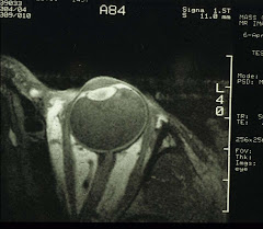
Maybe this is a good time to look at the size of human eyeballs. We have been able to convince some patients to volunteer for MR imaging of the eye:

Table I. Refractive error of patients and volunteers (5 eyes/group).

Type of Eye

Mean ± SD (diopter)

Range (diopter)

Nanophthalmos (phakic)

+10.42 ± 6.22

+4.50 – +18.50

Hyperopia

+3.42 ± 0.66

+3.00 - +4.50

Emmetropia

+0.21 ± 0.25

-0.25 - +0.50

Myopia

-10.25 ± 4.31

-6.25 - -15.50


Notice the nanophthalmic eyes all have very high hyperopia which in fact is one of the diagnostic indices.

And from MR images, it is possible to calculate the size of the eyeballs in 3D:

Table II. Ocular dimensions (mean ± SD mm) determined from MR images.

Type of Eye

Axial1

Equatorial1

Coronal(H)

Coronal(V)

Nanophthalmos

18.1 ± 2.02

19.0 ± 2.62

19.3 ± 3.02

18.2 ± 2.52

Hyperopia3

23.1 ± 1.0

24.1 ± 0.5

23.8 ± 0.3

23.3 ± 1.4

Emmetropia

22.9 ± 1.3

23.9 ± 1.2

23.9 ± 1.5

23.0 ± 1.9

Myopia4

28.5 ± 1.0

27.1 ± 1.5

28.3 ± 0.5

27.4 ± 0.7

1The equatorial lengths are significantly greater than the axial lengths: paired t-test P=0.006 and 0.025 for emmetropia and hyperopia, respectively. For nanophthalmic eyes, P=0.1 and for myopia, P=0.2.
2
Unpaired t-tests between nanophthalmos and emmetropia in all dimensions, P<0.02.

3No difference between hyperopia and emmetropia.
4Unpaired t-tests between myopia and emmetropia in all dimensions P<0.01.

Clearly, myopic eyes are big. And in contrast, nanophthalmic eyes are much smaller which is the hallmark of nanophthalmos.

If we look at the anterior segment closely:

Table III. Anterior segment dimensions (mean ± SD mm).

Type of Eye

Ant. Chamber Depth

Lens Thickness

Lens Diameter

Nanophthalmos

3.0 ± 0.11

5.3 ± 0.82

8.5 ± 0.4

Hyperopia3

3.1 ± 0.3

4.6 ± 0.6

8.7 ± 0.4

Emmetropia

3.0 ± 0.2

5.0 ± 0.8

8.4 ± 0.4

Myopia4

2.6 ± 0.6

4.0 ± 0.3

9.4 ± 1.3

1Unpaired t-test when calculated as % axial length between nanophthalmos (16.5±2.5) and emmetropia (13.0±1.1): P=0.025.
2
Unpaired t-test when calculated as % axial length between nanophthalmos (29.5±7.2) and emmetropia (21.7±2.9): P=0.06.
3
No significant difference between hyperopia and emmetropia.
4
No significant difference between emmetropia and myopia.

Hmm, the anterior chamber depth and the lens size in nanophthalmos are both similar to normal emmetropic and hyperopic eyes. So what else has gone wrong?

Table IV. Choroidal and scleral thickness (mean ± SD mm).


Axial

Vertical

Equatorial

Type of Eye

Choroid

Sclera

Choroid1

Sclera

Choroid

Sclera2

Naophthalmos

0.7±0.3

0.7±0.3

0.7±0.3

0.5±0.2

0.6±0.3

0.7±0.3

Hyperopia3

1.0±0.0

0.7±0.3

0.4±0.2

0.4±0.2

0.5±0.3

0.4±0.3

Emmetropia

0.9±0.2

0.6±0.3

0.5±0.1

0.4±0.2

0.5±0.2

0.4±0.2

Myopia4

N.M.

N.M.

N.M.

N.M.

N.M.

N.M.

1Unpaired t-test between nanophthalmos and emmetropia: P<0.09; when calculated as % of total vertical axial length, nanophthalmos (4.0±1.8) vs. emmetropia (1.9±0.5): P<0.04. 2Unpaired t-test between nanophthalmos and emmetropia: P<0.08; when calculated as % of total equatorial axial length, nanophthalmos (3.4±1.4) vs. emmetropia (1.6±0.5): P=0.03. 3No difference between hyperopia and emmetropia. 4N.M. - Not measurable. Thicknesses less than the in-plane resolution of 0.31mm.

Ah, so that's it: the equatorial sclera is the thickest in the nanopthalmic eyes. It is almost like all eyes are given the same amount of building materials. Smaller eyes are constructed with thicker walls while larger (myopic) eyes with thinner walls. Thick equatorial sclera chokes off blood circulation through the vortex veins (each eye has 4 or 6 of these). The only effective treatment for relieving high eye pressure is therefore sclerostomy around the base of the vortex veins.

For those who have patiently read through the above, a bonus for you. We'll now show you how to perform high-resolution MRI of the eye (also the methodology used in the study described above):

MR imaging was conducted using General Electric 1.5 Tesla Signa body scanners. A home-made 1.25-inch receive-only surface coil tuned to the proton frequency of the imagers was placed over the eye of interest. [Note: the factory 3-inch TMJ coil also can be used.] The coil was constructed with variable capacitors that allowed tuning and matching. A sagittal head image series using an FOV of 20 mm or more was acquired to locate the eye. Slice selection in the axial plane was then determined using graphic prescription. Multi-slice images in the axial and coronal planes were obtained (usually 8-9 slices/plane/eye) using a T1-weighted spin-echo sequence with for example, TR/TE = 400/20 msec with an image resolution of 0.31 x 0.31 mm and a slice thickness of 3 mm (in this case, the image acquisition time was 4.5 min). Image slices was selected by aligning imaging planes as parallel to the antero-posterior axis as possible.