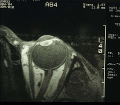
First, a quick review of the structure of the vitreous. The vitreous occupies the bulk (80%) of the eyeball. It is 99% water and has a lattice of collagen fibers with coils of hyaluronic acid distributing within the structure. Essentially, the vitreous is an optically clear gel. In the aging eye, parts of the vitreous become liquefied, and the collagen fibrils condense to form the floaters.
An example of this type of floaters is shown below:
 Notice the tadpole-shaped shadow in the 2 o'clock position. Cataracts also cast small shadows which are more centrally located, i.e., in the macular area. Floaters are more noticeable if they are situated close to the retina. However, not all intra-vitreous opacities are perceived by the patients. A good example is the tiny soap-like particles known as the asteroid hyalosis:
 These particles usually appear in only one eye. Sometimes it is an indication of (often undiagnosed) diabetes, so a blood glucose test is recommended.
Sudden onset of floaters can be a sign of trouble. A good example is tractional retina detachment (RD) during which, a blood vessel is ripped open. The blood cells appear as a sudden shower of floaters when they enter the vitreous. Retinal detachment requires surgical repair, that in itself is an entire branch of ophthalmology. An image of RD is shown below:
 (This one image courtesy of Optos)
 Again, the image may look like a total mess, the central vision is actually preserved.
Then we have vitreous hemorrhage from a leaking artery:
 Notice the origin of the hemorrhage (the dark red spot) is next to the optic disc in the 5 o'clock position. This hemorrhage actually extends into the vitreous seen by the patient as a large floater. A stated before, extensive vitreous hemorrhage requires vitrectomy in order to regain vision.
Finally, a very common condition known as posterior vitreous detachment (PVD). PVD affects 50% of people over 50. Typically the patient sees a sudden appearance of flashes together with a long curvilinear or a circular floater. Sometimes PVD is accompanied by hemorrhage and/or RD, but mostly quite benign except for the annoying floater. One such is shown below:
 This circular floater (which appears oval, a sideway view) is initially part of the vitreous attachment to the optic disc, in other words, a rim, which is pulled off during the vitreous separation from the retina. Luckily, no blood vessels are disrupted this time. Once the traction is relieved, the flashes are no longer seen.
So sudden onset of floaters must be treated as an emergency and the etiology identified.