Sunday, March 2, 2008

7.26 Eye-tooth, tooth-eye

News headline: "Blind Man Regains Sight After Doctors Implant Son's Tooth in His Eye" (Foxnews.com, 28 Feb, 2008)

So what is this all about? Well, there are always surgeries of the last-resort in any field.

As far as the eye, it is always a shame when only the cornea is messed up while the rest of the eye is intact. The logical next step is corneal transplant, typically around 90% success. What about the other 10%, though? They still need to see, don't they? Luckily, there are always surgeons who are willing to tinker, explore, invent, experiment, and cure. We will cite a few examples in this post.

Before we did that, a quick review on what could cause the corneal grafts to fail. Not surprisingly, the major one is allograft rejection, followed by increased intraocular pressure, infection (excluding endophthalmitis), and ocular surface problems. Pre-existing conditions such as diabetes and glaucoma increase the endothelial cell loss hastening the demise of the transplanted cornea. And chemical burn and end-stage dry eye (the latter as part of, e.g., Stevens-Johnson syndrome) both involving ocular surface changes also are major factors in graft failure. Any of the above leads to undesirable outcome, even multiple corneal transplants would not take.

The alternative is then to implant optically active prosthetics directly into the cornea. And if the retina is functioning well, then the patients can regain functional vision often at 20/40 or better.

First example is the Boston Keratoprosthesis developed by Dr Claes Dohlman of Massachusetts Eye and Ear Infirmary in Boston (a schematic is shown below):

The locking ring is titanium, it therefore does not interfere with MRI. This device is first fitted into a donor cornea, then the whole assembly is transplanted as that in normal penetrating keratoplasty. And a therapeutic contact lens is then fitted over the implant, together with life-long use of antibiotic eyedrops. The outcome is quite dramatic, interested readers can look up a news article "No time for tears" in the 5 Nov 2007 issue of Boston Globe.

The post-op appearance (without the contact lens) is shown below:

The second example is OOKP (osteo-odonto keratoprosthesis) developed by the late Dr GianCarlo Falcinelli of San Camillo Hospital in Rome. In patients with severe dry eyes, Boston Keratoprosthesis may not perform well owing to the need for a contact lens - hence the need for adequate tear fluid production. These patients will have to have an eye-tooth implant. Eye-tooth = the upper canine tooth because it situates near the eye. Only a few surgeons in the world are qualified to perform this procedure that include Drs Christopher Liu of Brighton, England, Günther Grabner in Salzburg, Austria, and Konrad Hille of Homburg/Saar, Germany. And a team led by Dr Donald Tan of Singapore National Eye Centre is also active in OOKP implantation. Other eye centers may have also followed suit, check your local listing.

OOKP implant is a two-stage process. The first involves the repair of ocular surfaces using mucosal linings from the patient's cheek, removal of a canine tooth with part of the jaw bone, the tooth is fashioned into a bolt-shaped structure or a flat lamina with a hole drilled in the center, an optical cylinder is then inserted and cemented in the hole. The whole assembly is then implanted in the patient's cheek or under the fellow eye to allow growth of blood vessels. The second stage involves removal of anterior ocular contents and finally replacement with the tooth-bone-cylinder complex, now the tooth-eye.

Not all patients are surgically eligible for the procedures described above. When done, these last-ditch efforts often yield miracle-like results. These surgeons are a special lot, so are the patients who often have already endured multiple surgeries.

Wednesday, February 27, 2008

7.25 Gee, I feel faint

(Geronimo Mercuriale, 1530-1606 -from www.sahha.gov.mt)

In 1580, Geronimo Mercuriale formulated the concept of syncope and demonstrated that it was associated with a slow pulse rate: "Ubi pulsus sit rarus semper expectanda est syncope".

The precise cause of syncope, commonly known as fainting, is hard to pin down. The mechanisms, however, are quite well-known, for at least a few hundred years. Essentially, people faint because of a mixture of (1) cardiac inhibition from enhanced parasympathetic tone, and (2) vasodilation from sympathetic withdrawal. Simply put, a slowed heart rate that reduces blood flow to the brain thereby causing a patient to faint.

Like migraine headaches, there are many triggers for syncope - officially "vasovagal syncope", for example, stress, standing upright for too long [known to happen to the Guards at the Buckingham Palace], pain, the sight of blood, hunger, prolonged hyperthermia, just to name a major few. For a few seconds, the patient feels light-headed, experiences nausea, sweating, ringing in the ears, and visual disturbances, before losing consciousness. The patient then keels over and this is when the injuries occur.

(Changing of guards - before somebody fainted.)

(Avoiding the sight of blood, Mr Patroclus?)

Feeling faint in an eye doctor's office? Of course that happens. This is often confused with feeling dizzy or having blackouts; both of which need to be neurologically evaluated. And it is not quite the "true syncope" but the "near syncope". In other words, the patient has all the signs and symptoms but stops short of passing out. This occurs usually from four activities: (1) contact tonometry (both applanation and indentation - the instruments must touch the cornea to get IOP readings); (2) first-time contact lens trial; (3) slit-lamp biomicroscopy; and (4) insertion of punctal plugs. In all situations, once the patient's eyes or eyelids are touched by the tip of the tonometer, by the doctor or an assistant when inserting a contact lens, or by the doctor's pulling down the patient's lower eyelid for a closer view of the palpebral conjunctiva or the puncta, usually at the slit-lamp, vasovagal reflex kicks in within minutes. Interestingly, most patients are young and healthy, possibly for the first time in their lives, their eyes are now manipulated by a stranger. The eyes/eyelids are indeed under some mechanical pressure albeit quite minor yet the physical reaction is quite dramatic.

First aid is to have the patient lie down while raising his/her feet or at least lower the head, so the brain can get back some much-needed blood.

Call an ambulance? Nah. The patient recovers on his/her own within minutes. Although it is not unheard of that a complete cardiac work-up is ordered if the patient ends up in the ER. And at least one patient brought a lawsuit against one eye doctor for some unspecified damages and emotional stress (remember: this is the United States of America, home to many personal injury barristers).

Some patients are disappointed that they probably won't be able to wear contacts, ever. Some are a bit embarrassed yet are quite happy to have a tale for their loved ones. Fortunately, vasovagal syncope, true or near, is never fatal.

Think about this: For patients with a history of having trouble instilling eyedrops or inserting contacts, it may not be a matter of dexterity but a mild form of vasovagal reaction.

Sunday, February 24, 2008

7.24 You have small eyes

(A General Electric 1.5 Tesla Signa MR scanner with a head coil inside the bore)

Enough fun with the Singapore sling, now we move on to something intriguing.

Despite the outward appearance, all emmetropic and hyperopic eyeballs are pretty much the same in size. The myopic eyes are indeed a little bigger. And once in a while, we need to inform a patient, "You have small eyes." This is not being impolite, but to point out the presence of nanophthalmos (dwarf eye). Nanopthalmos is not to be confused with microphthalmia (small eye); the latter is a congenital malformation with other developmental issues, often from fetal alcohol syndrome or viral infection in utero. Patients with hereditary nanophthalmos may have a defective gene on Chromosome 11; otherwise, they are developmentally normal.

Nanophthalmos with uveal effusion is a nasty condition first described by Dr Robert Brockhurst of Boston, Massachusetts, in 1974. These patients often develop narrow-angle glaucoma that prompted surgical intervention. Unfortunately, this also results in secondary retinal and choroidal detachment - owing to uveal effusion. Further repair is ineffective and sometimes choroidal elevation can be mistaken as intraocular tumors. So the recognition of nanophthalmos is important in order to manage it correctly.

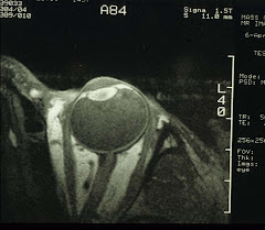
Maybe this is a good time to look at the size of human eyeballs. We have been able to convince some patients to volunteer for MR imaging of the eye:

Table I. Refractive error of patients and volunteers (5 eyes/group).

Type of Eye

Mean ± SD (diopter)

Range (diopter)

Nanophthalmos (phakic)

+10.42 ± 6.22

+4.50 – +18.50

Hyperopia

+3.42 ± 0.66

+3.00 - +4.50

Emmetropia

+0.21 ± 0.25

-0.25 - +0.50

Myopia

-10.25 ± 4.31

-6.25 - -15.50


Notice the nanophthalmic eyes all have very high hyperopia which in fact is one of the diagnostic indices.

And from MR images, it is possible to calculate the size of the eyeballs in 3D:

Table II. Ocular dimensions (mean ± SD mm) determined from MR images.

Type of Eye

Axial1

Equatorial1

Coronal(H)

Coronal(V)

Nanophthalmos

18.1 ± 2.02

19.0 ± 2.62

19.3 ± 3.02

18.2 ± 2.52

Hyperopia3

23.1 ± 1.0

24.1 ± 0.5

23.8 ± 0.3

23.3 ± 1.4

Emmetropia

22.9 ± 1.3

23.9 ± 1.2

23.9 ± 1.5

23.0 ± 1.9

Myopia4

28.5 ± 1.0

27.1 ± 1.5

28.3 ± 0.5

27.4 ± 0.7

1The equatorial lengths are significantly greater than the axial lengths: paired t-test P=0.006 and 0.025 for emmetropia and hyperopia, respectively. For nanophthalmic eyes, P=0.1 and for myopia, P=0.2.
2
Unpaired t-tests between nanophthalmos and emmetropia in all dimensions, P<0.02.

3No difference between hyperopia and emmetropia.
4Unpaired t-tests between myopia and emmetropia in all dimensions P<0.01.

Clearly, myopic eyes are big. And in contrast, nanophthalmic eyes are much smaller which is the hallmark of nanophthalmos.

If we look at the anterior segment closely:

Table III. Anterior segment dimensions (mean ± SD mm).

Type of Eye

Ant. Chamber Depth

Lens Thickness

Lens Diameter

Nanophthalmos

3.0 ± 0.11

5.3 ± 0.82

8.5 ± 0.4

Hyperopia3

3.1 ± 0.3

4.6 ± 0.6

8.7 ± 0.4

Emmetropia

3.0 ± 0.2

5.0 ± 0.8

8.4 ± 0.4

Myopia4

2.6 ± 0.6

4.0 ± 0.3

9.4 ± 1.3

1Unpaired t-test when calculated as % axial length between nanophthalmos (16.5±2.5) and emmetropia (13.0±1.1): P=0.025.
2
Unpaired t-test when calculated as % axial length between nanophthalmos (29.5±7.2) and emmetropia (21.7±2.9): P=0.06.
3
No significant difference between hyperopia and emmetropia.
4
No significant difference between emmetropia and myopia.

Hmm, the anterior chamber depth and the lens size in nanophthalmos are both similar to normal emmetropic and hyperopic eyes. So what else has gone wrong?

Table IV. Choroidal and scleral thickness (mean ± SD mm).


Axial

Vertical

Equatorial

Type of Eye

Choroid

Sclera

Choroid1

Sclera

Choroid

Sclera2

Naophthalmos

0.7±0.3

0.7±0.3

0.7±0.3

0.5±0.2

0.6±0.3

0.7±0.3

Hyperopia3

1.0±0.0

0.7±0.3

0.4±0.2

0.4±0.2

0.5±0.3

0.4±0.3

Emmetropia

0.9±0.2

0.6±0.3

0.5±0.1

0.4±0.2

0.5±0.2

0.4±0.2

Myopia4

N.M.

N.M.

N.M.

N.M.

N.M.

N.M.

1Unpaired t-test between nanophthalmos and emmetropia: P<0.09; when calculated as % of total vertical axial length, nanophthalmos (4.0±1.8) vs. emmetropia (1.9±0.5): P<0.04. 2Unpaired t-test between nanophthalmos and emmetropia: P<0.08; when calculated as % of total equatorial axial length, nanophthalmos (3.4±1.4) vs. emmetropia (1.6±0.5): P=0.03. 3No difference between hyperopia and emmetropia. 4N.M. - Not measurable. Thicknesses less than the in-plane resolution of 0.31mm.

Ah, so that's it: the equatorial sclera is the thickest in the nanopthalmic eyes. It is almost like all eyes are given the same amount of building materials. Smaller eyes are constructed with thicker walls while larger (myopic) eyes with thinner walls. Thick equatorial sclera chokes off blood circulation through the vortex veins (each eye has 4 or 6 of these). The only effective treatment for relieving high eye pressure is therefore sclerostomy around the base of the vortex veins.

For those who have patiently read through the above, a bonus for you. We'll now show you how to perform high-resolution MRI of the eye (also the methodology used in the study described above):

MR imaging was conducted using General Electric 1.5 Tesla Signa body scanners. A home-made 1.25-inch receive-only surface coil tuned to the proton frequency of the imagers was placed over the eye of interest. [Note: the factory 3-inch TMJ coil also can be used.] The coil was constructed with variable capacitors that allowed tuning and matching. A sagittal head image series using an FOV of 20 mm or more was acquired to locate the eye. Slice selection in the axial plane was then determined using graphic prescription. Multi-slice images in the axial and coronal planes were obtained (usually 8-9 slices/plane/eye) using a T1-weighted spin-echo sequence with for example, TR/TE = 400/20 msec with an image resolution of 0.31 x 0.31 mm and a slice thickness of 3 mm (in this case, the image acquisition time was 4.5 min). Image slices was selected by aligning imaging planes as parallel to the antero-posterior axis as possible.

Sunday, February 17, 2008

7.23 Singapore sling

Please, Singapore is pronounced Sin-ga-po', not Sin-ga-poah. And Singapore sling is a "mysteriously refreshing drink" in the tropical sun, not a run-of-the-mill cocktail at your local pub. There are many imitations, though. For DIYers, this one, from drinkmixer.com, is quite genuine:

1 oz gin
1/2 oz cherry brandy
4 oz pineapple juice
1/2 oz lime juice
1/4 oz Cointreau® orange liqueur
1/4 oz benedictine herbal liqueur
1/3 oz grenadine syrup
1 dash Angostura® bitters
Combine all in a shaker, fill with ice and shake until the shaker is well frosted. Strain into a tall glass and garnish with a slice of pineapple and cherry. No tiny umbrellas of course.

Better yet, go down to the Long Bar at Raffles Hotel, sit in the rattan wicker chair and have the sling with roasted-in-shell peanuts. You'll forget where you are.

Ah, but we do remember: Singapore is a prefect place to investigate the pattern of cataract formation. The Merlion City is sitting practically on the equator (1°N) with 12 hours each of day and night, with an annual temperature average of 26.7°C and a UV index of 10-13. These couldn't be good for the eyes? Yet, sunglasses and broad-rimmed hats are apparently not in fashion (see image below).

See, only one guy with sunglasses and none wears a hat.

The Merlion? Here-lah:

So, in a study on 550 Chinese Singaporeans of older than 50 years, only 26.1% are found without cataracts. More specifically, 47.3% of the 50-60 age group are without cataracts; although the rate drops down to 11.7% in the 60-70 group. And everybody has cataracts if older than 70.

Compared with a group of Japanese (884 participants) residing in Noto Peninsula - which has an average temperature of 21.3°C with 1/2 the UV radiation of S'pore - cataract prevalence in the 50-60 Singaporeans is similar to that of the 70-80 age group in Japan, i.e., a decade earlier. In addition, nuclear cataract prevalence in the 50-60 Singaporeans was 9 times higher than the same age group in Japan. One can argue that besides geography, there are differences in culture, e.g., diet, outdoor activities, etc that may also be contributing factors. The principal factors are, however, still ambient temperature and UV index.

In fact, in cooler places like Iceland, with an average temperature of 4.4°C and 1/5 of UV index of S'pore, 39% of 60-70 year-olds are still cataract-free (study population=993). If race is a major factor, then all Caucasians no matter where they live should have identical cataract prevalence. This is not the case: Melbourne residents in fact have higher cataract prevalence than Icelanders.

Further, the prevalence of nuclear cataracts in Sumatra, another high temperature locale, is similar to that of S'pore suggesting the high ambient temperature, not race is a major factor.

Does it really matter to have high cataract prevalence? It does from the viewpoint of loss of productivity and the costs for cataract extraction. It is much simpler to practice prevention - clearly a job for the eye doctors.

Have another sip and plan.

(A more detailed version of this post, written in Chinese, is available here.)

Friday, February 15, 2008

7.22 One, two, or three eyes

(Yang Jian, the 3-eyed god-general)

In Homer's Odyssey, Book 9, Mr Odysseus ran into Mr Polyphemus, a cyclops. The one-eyed giant ate a few of Odysseus's crew before losing his eye to Mr O's trickery. In Chinese mythology, The Investiture of the Gods (封神演義), Chapter 97, Yang Jian (楊戩) was born with three eyes; the third, sitting vertically on the forehead, can see beyond space and time.

While it is possible for a vertebrate embryo to develop only one eye, the three-eyed variety probably does not exist because this will require a fundamental re-structure of the cerebrum into three 1/3 brains. If we want to venture a guess, General Yang's third eye was most likely either a birthmark or a nevus that looked like an eye.

Cyclopes result from a problem with the prosencephalon which normally divides into right and left brains. Failing so, only one eye forms in the place where the nose is supposed to be. Cyclopes are mostly stillborns, so it was highly unusual for Polyphemus to even grow up, but then his Dad probably intervened (as far as life-support).

Actually, a close inspection reveals a complicated origin for each tissue in the eye. For those who for some reason must know, here is a quick summary:

Neuroectoderm: the retina, epithelium of the ciliary body/iris, and optic nerve
Surface ectoderm: the lens and corneal epithelium
The surrounding mesenchyme (neural crest origin): the sclera, the remainder of the cornea, the choroid, ciliary body/iris, and blood vessels

In the vertebrates, the development of the eye is through several invagination processes with precise timing. Any insults from microbes or toxins can of course cause mutations and congenital eye diseases. (See previous posts under Pediatrics.)

Here is something new and interesting: The development of the lens maybe controlled by the Six3 gene which in turn regulates Pax6, the master gene for eye development. The presence of gene product of Six3 does precede that of Pax6. In fact, problem with Six3 leads to holoprosencephaly. And the worse case? Yes, Mr Polyphemus the Cyclops and a kitten named Cy (see image below).

(Cy the kitten, the only living cyclops ever, even if for only one day - from cbsnews.com)

So Six3, in effect, initiates the whole cascade of gene activation in the development of the eye. We wonder why Poseidon did not see this coming.

Tuesday, February 12, 2008

7.21 Never say never?

(Warner Bros, 1983)

After years of education by sci-fi movies and TV shows, some members of the general public actually believe eye transplant is a reality. Well, it is not, not yet anyway. And is it just a pipe dream or shall we quote Mr James Bond, "Never say never again" in view of the tremendous progress in recent medical research?

If it is possible to transplant anything at all, the candidates must have been blinded by an incurable eye disease yet still with some residual, functional neural (visual) activity. And the procedures must of course subject to the IRB approval.

Now, let's review what the major obstacles may be, one scenario at a time - an FAQ of sorts:

1. Transplant the whole eyeball?

Sure, assuming we can keep the donor eyeballs alive, or more important, for the tissues, especially the cornea, lens, and the retina, to remain functional after transplant. The eyeballs must be kept at 0-4°C; although the best way is probably to connect the arteries, branching from the ophthalmic artery, to a miniature circulator using oxygenated artificial blood enriched with 5.5mM glucose, so that the ocular group of arteries can continue to supply and sustain tissue metabolism.

Now we come to the second problem: how to reconnect the optic nerve.

Picture an undersea fiberoptic cable that got severed during a major earthquake. Do you then retrieve the cable, and reconnect the many thousands of fibers one by one? No, you'd replace the entire section. The optic nerve has 1 million nerve fibers. And at present, there is no way of joining two damaged nerve fibers to make it one functional unit. Also, unlike the undersea cables, the optic nerve is not an isolated section with connectors at both ends, so you cannot replace the optic nerve itself.

Assuming you can somehow coax the nerve fibers to repair their cell membranes and join end to end, it probably won't be the whole 1 million fibers. Then it depends on how many are revived which will then determine the visual field size and the attainable visual acuity.

After all the above, the visual cortex will need to exercise all its plasticity to interpret the images, since it now receives signals from most definitely mis-wired optic nerve fibers. Whether this is possible is still anybody's guess.

Last but not least, the extraocular muscles must be re-attached so that the eye can move according to the direction of gaze. This is cosmetically important as well.

So, transplanting the whole eyeball seems a bit impractical, at least for now.

2. Transplant the retina?

OK. The full-thickness retina or just the photoreceptor layer? The whole retina or just the macula?

First, the retinal viability and the reconnection of "wires" (this time with the optic nerve) are both still the major obstacles. The image below is a whole mount retina specially stained for the retinal ganglion cell fibers (downloaded from webvision.med.utah.edu). You can see numerous fibers all converging toward the optic disc (the black "hole" in the center) and each one must be re-connected with its corresponding fiber in the optic nerve. Technically, with open-sky vitrectomy, perhaps the whole thickness retina/macula can be replaced in toto, but not the photoreceptor layer itself (remember it faces posteriorly the pigment epithelium) - it is simply a humanly impossible task.

Still a bit unrealistic, this retinal transplant deal. But then as in eye transplant, it may just be a matter of figuring out how to fuse nerve fibers through some clever means. And other innovations may then follow.

3. What about electronics?

Ah, now we enter the realm of practicality. A project led by Dr Eberhart Zrenner of Tübingen and Regensburg universities in Germany has implanted retinal chips in patients with retinitis pigmentosa (see image below - from The Economist, 7 June, 2007).

This chip is composed of 1,540 photodiodes which, when stimulated by light, send electric pulses to retinal ganglion cells and the excitation is transmitted all the way to the visual cortex. So now we have roughly 40 x 40 pixels that cover most of the visual field. The patients can differentiate black and white as well as some shapes. For some, this is already a huge difference.

For patients without a semi-functional eye, the alternative is to implant electrodes directly into the visual cortex. The electrodes are connected to a spectacle-mounted digital camera which functions as the eye. As with retinal chips, the resolution is quite low (around 15 x 16 pixels) and the patients can only "see" white dots on black background.

These artificial sensors no doubt will further improve so that the resolution may become high enough with little power requirement and that each implanted chip can last a lifetime. There are also attempts to teach other senses such as auditory and taste to receive and interpret electronic visual signals - with varying degrees of success.

The electronics are not a bad first step towards bionic vision - a popular notion ever since the debut of the "Six Million Dollar Man" series (ABC TV) in 1978.

4. Stem cells?

Yes, this can be very promising especially for replenishing photoreceptor cells lost to retinal degeneration. Previous attempts of directly injecting retinal cells from young rabbits into the adult rabbit eyes have produced intriguing results: There was incorporation of the photoreceptors in the the retina that formed "rosettes". Photoreceptors were present at the luminal side of the rosettes surrounded by layers corresponding to inner layers of the normal retina. So the transplanted retinal "bits" appear to have a natural tendency to self re-organize. Stem cells grown into retinal cells, when implanted, will most likely behave the same way.

The potential problems in stem cell transplants are surgical injury, tumor formation, and vector-mediated infection. These can be avoided as the transplant process evolves. The biggest hurdle in stem cell research seems political, at least at the present time.

5. No more gene therapy?

Not at all. In fact, this research is proceeding in great earnest. As previously posted, gene therapy has already been tested in Leber's congenital amaurosis (LCA) to correct a defective RPE65 gene. This project was carried out last year at University College London Institute of Ophthalmology and Moorfields Eye Hospital, led by Prof Robin Ali with Drs James Bainbridge and Tony Moore. If successful, it will no doubt lead and change the way of genetic eye disease treatment.

Hmm, James Bond maybe right after all.

Monday, February 11, 2008

7.20 Mr Sulu, take us home

(Star Trek IV, The Voyage Home, 1986)

You all remember the Star Trek movie, "The Wrath of Khan (1982)", in which Captain James Tiberius Kirk receives a pair of reading glasses as a birthday present from Dr Leonard (Bones) H McCoy. Kirk proceeds to lament rather unconvincingly the onslaught of old age. Presumably, by the 23rd Century, presbyopia has already been eradicated. Unfortunately, Capt Kirk is allergic to Retinax V - the medicine for treating Presbyopia, or "老花眼old blurry eyes" in Chinese and "老眼old eyes" in Japanese. The keyword: "old".

Funny the script did not follow the more creative vein as that for Captain Jean-Luc Picard who chooses to be bald (Ha!!) There is perhaps a lack of imagination on the screen writers' part. Because even now, there are invasive ways of manipulating presbyopia. Not that this is necessary, a pair of inexpensive OTC readers, bifocals including bifocal contacts, or monovision correction of various kinds (from LASIK to contact lenses, to spectacles) all can quickly resolve the issue.

Presbyopia is an age-related loss of crystalline lens deformability for focusing at near. This was described by Thomas Young (1773-1829) in 1793. To explain the process, he had invoked the elasticity theory. The Young's Modulus (stress = E x strain) is dependent on the material. In the case of accommodation, the lens capsule appears the candidate (see image below). Young's argument was that the capsule elasticity had diminished during aging. Indeed, recent measurements confirmed that the Modulus was about 6 × 10⁷ dyn/cm2 in children which decreased to 3 × 10⁷ dyn/cm2 at age 60, and to 1·5 × 107 dyn/cm2 in extreme old age. Similar to tired old rubber bands which cannot resume the original lengths when the tension is released.

(Lens capsule is the outer most layer which envelops the whole lens
- it is the faint band to the left (anterior to) the single-cell-layer epithelium in the above image)

That is not the whole story, though. There are three parts involved in the accommodation process (see below, 1-3):
(1: The zonules; 2: the crystalline lens; and 3: ciliary muscle)

The ciliary muscle must first contract to allow a decrease in the tension of the zonules, so the capsule can re-shape the lens. Does the ciliary muscle change with aging as well? Not the contractility. It appears the diameter of the ciliary muscle ring does decrease owing to a configurational change. The overall result is the zonules no longer have enough space to relax. And the weakened capsule also can no longer deform the lens as much as before. Did the lens diameter increase with aging also? Some say no; although the only way to make sure is to do a large-scale high-resolution MRI of the eye, because the lens is hiding behind the iris, inaccessible through optical means. The zonules? It is assumed, but never quantified, that the zonular fiber elasticity also changes with time.

Loose ends galore, indeed.

So, how can presbyopia be manipulated surgically? At least in theory, the ciliary muscle ring can be re-positioned to create more space for the zonules. This procedure has already been attempted; although the outcome is still unclear. Alternatively, Conductive Keratoplasty (CK), using radiofrequency to heat and shrink collagen fibers in the cornea, can be done. This to steepen the curvature of the appropriate area of the cornea, so that more plus power is produced for near tasks. CK takes about 3 min and is done to only one eye - a monovision correction as well. It seems to work for some, for a limited duration anyway (about 4-7 years). Interested parties are urged to consult their own eye doctors.

It's been 26 years since "The Wrath of Khan" and we still don't know the composition and the mechanism of effect of Retinax. Well, the sci-fi writers have already pointed the way, perhaps pharmacological researchers can now take us home - to a presbyopia-free world.