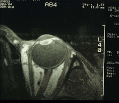
Unfortunately in the young eyes, we can find older people's problems, e.g., cataract and glaucoma. Congenital cataract and congenital glaucoma, that is. By far, these are the two major congenital anomalies of the eye.
There are inherent difficulties in examining the eyes of a tiny infant. A good example is the visual acuity which cannot be assessed accurately. Yet another is the measurement of the intraocular pressure. And often sedation is needed in order to perform a complete exam, and which often must be done in an OR setting. Refraction, on the other hand, can be done with trial-lens retinoscopy or a hand-held auto-refractor. The latter, however, is less useful if nystagmus is present. And contact lens fitting naturally requires full parental participation. Routine interactive tests, e.g., subjective refraction and visual field testing must be postponed until much later for obvious reasons.
Congenital cataract is diagnosed at birth or it can develop soon after. In 1/3 of the cases, cataract is present in only one eye. If in both eyes, then 23% of the patients have a family history in an autosomal dominant pattern. This type of congenital cataracts is frequently associated with metabolic/systemic diseases (e.g., hypolycemia, trisomy, and myotonic dystrophy). Some congenital cataracts are a result of infection in-utero, most commonly from rubella; although it can also be from a host of others including rubeola, chicken pox, cytomegalovirus, herpes simplex/zoster, poliomyelitis, influenza, Epstein-Barr virus, syphilis, and toxoplasmosis. In under-developed countries, it can be from poisons in the drinking water contaminated by industrial wastes.
Because of the early presentation, if the opacity obstructs vision and left untreated, amblyopia can develop in the affected eye, and permanent vision loss if both eyes are cataractous. The usual guideline is if the opacity is 3mm or greater and located in the path of the visual axis, then the lens must be extracted. Smaller opacities do not necessarily cause vision issues. They are often discovered by chance during an adulthood routine eye exam, to the patient's greatest surprise.
Congenital glaucoma is also present at birth; although most cases are detected during early infancy/childhood. It is caused by a malformation in the fluid drainage channels, known as the trabecular meshwork, in the eye. Very rarely it is hereditary; although it won't be surprising if some cases are. It can affect only one eye; however, in 70% of the cases, both eyes. And more in boys (65%). The increase in the intraocular pressure from fluid build-up can rupture the corneal endothelium causing entry of water into the cornea. And the eye itself enlarges in size as well. Like glaucoma in the adults, the retina can be permanently damaged.
Congenital glaucoma is treated with surgical creation of a drainage pathway. Often multiple surgeries are needed to finally stablize the intraocular pressure. As you can imagine, this requires the expertise of a pediatric ophthalmologist specializing in congenital glaucoma. A video from the University of Iowa demonstrating trabeculotomy is shown below:
http://webeye.ophth.uiowa.edu/eyeforum/cases/case42-Primary-Congenital-
Glaucoma-(Infantile-Glaucoma).htm
The eye is of course only a small part of the body which, while still in the developmental stage in the uterus, is subject to all sorts of assaults. Proper prenatal care, a healthy pregnancy, and full-term birth, can certainly go a long way towards avoiding all congenital diseases, not just cataract and glaucoma.
Glaucoma-(Infantile-Glaucoma).htm


No comments:
Post a Comment