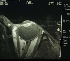
If it is possible to transplant anything at all, the candidates must have been blinded by an incurable eye disease yet still with some residual, functional neural (visual) activity. And the procedures must of course subject to the IRB approval.
Now, let's review what the major obstacles may be, one scenario at a time - an FAQ of sorts:
1. Transplant the whole eyeball?
Sure, assuming we can keep the donor eyeballs alive, or more important, for the tissues, especially the cornea, lens, and the retina, to remain functional after transplant. The eyeballs must be kept at 0-4°C; although the best way is probably to connect the arteries, branching from the ophthalmic artery, to a miniature circulator using oxygenated artificial blood enriched with 5.5mM glucose, so that the ocular group of arteries can continue to supply and sustain tissue metabolism.
Now we come to the second problem: how to reconnect the optic nerve.
Picture an undersea fiberoptic cable that got severed during a major earthquake. Do you then retrieve the cable, and reconnect the many thousands of fibers one by one? No, you'd replace the entire section. The optic nerve has 1 million nerve fibers. And at present, there is no way of joining two damaged nerve fibers to make it one functional unit. Also, unlike the undersea cables, the optic nerve is not an isolated section with connectors at both ends, so you cannot replace the optic nerve itself.
Assuming you can somehow coax the nerve fibers to repair their cell membranes and join end to end, it probably won't be the whole 1 million fibers. Then it depends on how many are revived which will then determine the visual field size and the attainable visual acuity.
After all the above, the visual cortex will need to exercise all its plasticity to interpret the images, since it now receives signals from most definitely mis-wired optic nerve fibers. Whether this is possible is still anybody's guess.
Last but not least, the extraocular muscles must be re-attached so that the eye can move according to the direction of gaze. This is cosmetically important as well.
So, transplanting the whole eyeball seems a bit impractical, at least for now.
2. Transplant the retina?
OK. The full-thickness retina or just the photoreceptor layer? The whole retina or just the macula?
First, the retinal viability and the reconnection of "wires" (this time with the optic nerve) are both still the major obstacles. The image below is a whole mount retina specially stained for the retinal ganglion cell fibers (downloaded from webvision.med.utah.edu). You can see numerous fibers all converging toward the optic disc (the black "hole" in the center) and each one must be re-connected with its corresponding fiber in the optic nerve.
 Technically, with open-sky vitrectomy, perhaps the whole thickness retina/macula can be replaced in toto, but not the photoreceptor layer itself (remember it faces posteriorly the pigment epithelium) - it is simply a humanly impossible task.
Still a bit unrealistic, this retinal transplant deal. But then as in eye transplant, it may just be a matter of figuring out how to fuse nerve fibers through some clever means. And other innovations may then follow.
3. What about electronics?
Ah, now we enter the realm of practicality. A project led by Dr Eberhart Zrenner of Tübingen and Regensburg universities in Germany has implanted retinal chips in patients with retinitis pigmentosa (see image below - from The Economist, 7 June, 2007).
 This chip is composed of 1,540 photodiodes which, when stimulated by light, send electric pulses to retinal ganglion cells and the excitation is transmitted all the way to the visual cortex. So now we have roughly 40 x 40 pixels that cover most of the visual field. The patients can differentiate black and white as well as some shapes. For some, this is already a huge difference.
For patients without a semi-functional eye, the alternative is to implant electrodes directly into the visual cortex. The electrodes are connected to a spectacle-mounted digital camera which functions as the eye. As with retinal chips, the resolution is quite low (around 15 x 16 pixels) and the patients can only "see" white dots on black background.
These artificial sensors no doubt will further improve so that the resolution may become high enough with little power requirement and that each implanted chip can last a lifetime. There are also attempts to teach other senses such as auditory and taste to receive and interpret electronic visual signals - with varying degrees of success.
The electronics are not a bad first step towards bionic vision - a popular notion ever since the debut of the "Six Million Dollar Man" series (ABC TV) in 1978.
4. Stem cells?
Yes, this can be very promising especially for replenishing photoreceptor cells lost to retinal degeneration. Previous attempts of directly injecting retinal cells from young rabbits into the adult rabbit eyes have produced intriguing results: There was incorporation of the photoreceptors in the the retina that formed "rosettes". Photoreceptors were present at the luminal side of the rosettes surrounded by layers corresponding to inner layers of the normal retina. So the transplanted retinal "bits" appear to have a natural tendency to self re-organize. Stem cells grown into retinal cells, when implanted, will most likely behave the same way.
The potential problems in stem cell transplants are surgical injury, tumor formation, and vector-mediated infection. These can be avoided as the transplant process evolves. The biggest hurdle in stem cell research seems political, at least at the present time.
5. No more gene therapy?
Not at all. In fact, this research is proceeding in great earnest. As previously posted, gene therapy has already been tested in Leber's congenital amaurosis (LCA) to correct a defective RPE65 gene. This project was carried out last year at University College London Institute of Ophthalmology and Moorfields Eye Hospital, led by Prof Robin Ali with Drs James Bainbridge and Tony Moore. If successful, it will no doubt lead and change the way of genetic eye disease treatment.
Hmm, James Bond maybe right after all.


2 comments:
The information you provided was very helpful. You had wide coverage in small area. Thanks..
I am very much looking forward for the development in eye.
You are welcome.
After all the brain is just an extension of the eye. And yet more is known about the brain, hence my posts.
Per your request, I have added something about the development of the eye (see 7.22 One, two, or three eyes).
Have fun!!
Post a Comment