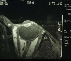
Fundamentally, we need to know if the ocular functions are normal. If not, are they related to structural abnormalities that can be observed optically. And if that is not possible, then imaging with either ultrasound or radiological means. Ultimately, a decision is made with regard to treatment.
So what are the functions that must be tested?
1. Visual acuity and contrast sensitivity
Visual acuity (VA) is a measure of resolution and contrast is the ability to grade a gray scale. Contrast sensitivity is rarely tested except in specialty services.
VA is a routine test for your vision at infinity. For all practical purposes, the distance between the patient and the acuity chart is set at 20 ft or 6m, not at infinity. The rationale is that at this distance, the amount of accommodation is only 1/6=0.17D. In theory, all results of refraction must correct for this 0.17D. However, since the spectacle lenses and contacts are available only in 0.25 steps, this correction is impractical and is therefore ignored.
So what does it mean when you have 20/20 vision? It means that on the VA chart, you can see the 20/20 line at 20 ft. A person with a 20/200 vision means this person can seen the line only at 20 ft while normal-sighted people can see it 200 ft away.
There are three types of VAs: uncorrected, pinhole, and best corrected. It is usually done one eye at a time, then both eyes together. Binocular vision is better than monocular because of the psychophysical summation (i.e., two guesses reinforce each other). Pinhole acuity is, as the name implies, your vision when bypassing the optical system of the eye. It is often very close to the bested correctable vision. However, if there is no improvement with the pinholes, then it is an indication of a problem in the visual pathway.
The denotation of VA varies from one continent to another. The US is probably the lone nation holding out on the Imperial system. The table below is a conversion chart between different systems:
Snellen Notation Metric Imperial | MAR | logMAR | Decimal | |
| 6/60 | 20/200 | 10 | 1.0 | 0.10 |
| 6/48 | 20/160 | 8.0 | 0.9 | 0.13 |
| 6/38 | 20/125 | 6.3 | 0.8 | 0.16 |
| 6/30 | 20/100 | 5.0 | 0.7 | 0.20 |
| 6/24 | 20/80 | 4.0 | 0.6 | 0.25 |
| 6/19 | 20/60 | 3.2 | 0.5 | 0.32 |
| 6/15 | 20/50 | 2.5 | 0.4 | 0.40 |
| 6/12 | 20/40 | 2.0 | 0.3 | 0.50 |
| 6/9.5 | 20/30 | 1.6 | 0.2 | 0.63 |
| 6/7.5 | 20/25 | 1.25 | 0.1 | 0.80 |
| 6/6 | 20/20 | 1.00 | 0.0 | 1.00 |
| 6/4.8 | 20/16 | 0.80 | -0.1 | 1.25 |
| 6/3.8 | 20/12.5 | 0.63 | -0.2 | 1.58 |
| 6/3.0 | 20/10 | 0.50 | -0.3 | 2.00 |
The Snellen chart is the chart with letters (optotypes) and the MAR charts are designed to reflect the minimum angle of resolution, often in a logarithmic form, or LogMAR.
The design of the VA chart is based on the subtended visual angle. For example, the letters of the 20/20 line of a projected Snellen chart is usually E V O T Z 2. Look at the E:
 Each "stroke" of the E subtends an angle of 1 min, or an arc on a circle with a radius of 20 ft (6m). This one-min arc seems the minimum for the retinal photoreceptors to resolve (within the confines of the optical system of the eye and the density of the cells). All other VA charts are based on the same principle.
Is it possible to see better than 20/20? Yes, in fact 20/15 is not unusual. And if the patient's eyes are designed for 20/15 vision, the endpoint of refractions cannot stop at 20/20 or the patient will still see blurred objects. The optical system of the eye is imperfect, it has a fairly large chromatic aberration. To see even better, we'll need the wavefront technology.
If we regard the light as a bundle of rays, then a line drawn perpendicular to these rays is called a wavefront. Which is flat when reaching an eye with no aberrations. Otherwise, it appears irregular. These (monochromatic) aberrations are the higher-order ones, such as coma, terfoil, spherical aberration, etc. Wavefront-guided LASIK is now the standard and indeed the vision can be corrected to better than 20/15. On the other hand, the optical application of wavefront technology, i.e., spectacles and contacts, is highly individual. And the gain probably does not justify the costs.
2. Cover and binocular tests
This is a simple test for the position of the eyes when they are dissociated - by covering one eye while the other looks (fixates) at an object. Then the cover is switched to the other eye, and the uncovered eye now re-fixates. If it moves nasally, that suggests the eye turned out when covered. This is a case of eso- phoria/-tropia. And so on and so forth.
To quickly find out if the two eyes work together, a simple Worth-4-dot test should do. The patient wears a John Lennon style specs with one green lens and the other red. The Worth-4-dots are 4 holes on the round glass plate of a flashlight (see picture below) with approximately 1cm in diameter for each hole. The holes are organized in a diamond shape. Conventionally, the top hole is red, both the left and right are green, and the bottom is white. If only one eye is functional and the other is suppressed, then the patient sees either 2 or 3 lights. If both eyes fixate together, then 4 lights. And if double vision, 5 lights.
To assess if stereopsis is present, a series of polarized images are shown to patients wearing polarized specs. The degree of stereo vision or even its absence can be readily tested.
And to see if all EOMs are working in coordination, by asking the patients to follow the movement of a penlight going horizontally, vertically, and obliquely, the EOM in trouble can be quickly identified.
3. Pupillary responses
This topic by itself can be the basis for a paperback. Let's bypass the neural pathways for now. We'll simply tabulate the abnormal pupillary responses ("+"=yes and "-"=no):
Pupil Anomalies | Anisocoria | Light reaction | Near reaction | 0.125 % pilocarpine | 4 % or 10% Cocaine | 1% hydroxy- amphetamine | 1 % pilocarpine |
Tonic pupil | (+) | (-) or minimal | (+) | (+) | No indication | No indication | No indication |
Acute third-nerve palsy | (+) | (-) | (-) | No indication | No indication | No indication | (+) |
Pharmacologic dilation | (+) | (-) | (-) | No indication | No indication | No indication | (-) |
Horner’s Syndrome | (+) | (+) | (+) | No indication | (-) | (+) 1st or 2nd order neurons | No indication |
Argyll Robertson pupils | (+) | (-) | (+) | No indication | No indication | No indication | No indication |
The pupil testing technique is knows as the swinging penlight test. Normally, the pupils of the two eyes are both round and equal in diameter. And when one eye is exposed to the bright penlight, its pupil and that of the other eye will both constrict. When the light is swung over to the other eye, both pupils will remain constricted. The pupils also constrict when looking from far to near. Anything else is not normal indicating a possible neurological deficit.
For example, if, when the penlight is swung over to the other eye, and the pupil, instead of remaining constricted, now dilates, then we have a Marcus-Gunn pupil. This suggests a lesion in the optic nerve.
And in the table above, anisocoria means unequal pupil size. Tonic pupil is usually a result of trauma. One particular form with unknown etiology, called Adie's tonic pupil, is fairly harmless often seen in young ladies. Argyll Robertson pupils are associated with neurosyphilis. And Horner's syndrome is often associated with cancer at the tip of the lungs that impinges upon the sympathetic nerve on the carotid artery.
The tests listed above are all low-tech ways that can be done easily. And they are in fact performed routinely in all clinics. The results can be quite informative, often alerting the doctors to the presence of potentially sight- or life-threatening diseases.


1 comment:
Usually I never comment on blogs but your article is so convincing that I never stop myself to say something about it. You’re doing a great job
Enjoyed reading the article above , really explains everything in detail,the article is very interesting and effective.Thank you and good luck for the upcoming articles. very interesting , good job and thanks for sharing such a good blog. I really appreciated to you on this quality work. Nice post!! these tips may help for all.
For Eye care and Eye treatment services visit
Smart Vision Eye Hospitals
Post a Comment