1. Auto-refractor: Most auto-refractors are now capable of both retinoscopy and keratometry. And within a few seconds, the values of both refractive error and corneal curvatures are obtained. The same data also can be acquired manually with a retinoscope and a keratometer, within a few minutes. So the time saved with the automated instrumentation may be important in a high volume practice. The quality of data is similar regardless of which way they are obtained.
On the other hand, far more data are generated with corneal topography than keratometry, the latter can examine only the central 3-4mm of the cornea and measure only the curvatures. Corneal topology, based on a reflection pattern of thousands of points of the entire cornea, is shown as a sagittal map. Which color-codes the curvatures according to their optical power. The data can be used for pre-LASIK planning as well as for fitting special contacts. And any corneal irregularities, e.g., keratoconus or distortion from scarring, can be easily seen.
2. Slit-lamp observation or biomicroscopy did not change much except for digital photographic attachments in newer models. Examination of the fundus with the slit-lamp and also that with either direct or indirect ophthalmoscopes can be done with a wide-field fundus camera, i.e., the Optomap retinal scanner (see picture below). Optomap imaging takes 0.25 sec/image and requires no pupil dilation. Subtle lesions, of course, can be further examined with dilated binocular indirect ophthalmoscopy.
In this case, the fast Optomap imaging for screening retinal lesions is far more convenient and comfortable for the patients. All primary care offices should consider using this instrument.
 3. Tonometry has not change much, either. It is either non-contact (air-puff), indentation, or applanation tonometry. Some instruments have hand-held models (an example is shown below) for cases with postural difficulties on the desktop models.
 4. Visual field examination can be done by using tangent screens or much more expeditiously with modern automated perimeters. The commonly used Humphrey visual field analyzers also come with a screening model. Which is based on the frequency doubling principle that can detect visual field loss due to M-cell neuron death (typically from glaucoma). And the examination can be done in a few minutes.
5. Retinal imaging has made great progress in the form of OCT (Optical Coherence Tomography). The image below is a good example:
 OCT is based on low coherence interferometry capable of sub-micron resolutions. It has two modes, time-domain and the more recent spectral-domain OCT. The latter is capable of ultra-high speed and ultra-high resolution, at 73 and 3 times that of the former, respectively. We will no doubt continue to witness the further progress of this technology.
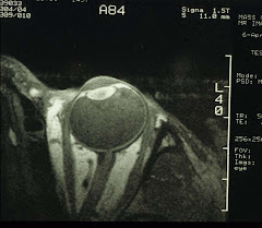
6. For gross anatomy of the eye, there is the old standby of office-based ultrsonography (in both A- and B-modes) and the MRI-based high-resolution surface coil imaging (see image on the rigt panel). X-Ray and CT scans for the eye are not recommended for the ionizing radiation that may cause cataracts. We favor the MRI approach as it gives far greater details than the ultrasound.
Obviously not every clinics is equipped with every piece of advanced instruments under the sun. Instruments, however, do not make diagnosis, doctors do.


No comments:
Post a Comment