 (The above is a fundus photo of optic neuritis, notice the blurred disc margin. This is not always seen, however. Image from meded.ucsd.edu.)
Sometimes a patient's chief complaint is this: Over the past few weeks, vision fades in and out and the patient is now experiencing "pain behind the eye" especially when looking about. Also, there is now a loss of vision. A quick visual acuity test does confirm the patient's own observation. And the swinging penlight test also reveals an afferent pupillary defect. Ophthalmoscopy, however, shows a normal optic disc. You are entertaining a diagnosis of optic neuritis but how to confirm it in the absence of any obvious signs of disc change?
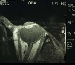
Contrast MRI, naturally:
 The above is a T1-weighted coronal image of the brain after intravenous GdDTPA, a contrast agent, to provide enhancement of the lesion. And in this case, a hyperintense left optic nerve (on the right side of the image, the white spot next to the caption) suggesting a leaky optic nerve sheath resulting from, most likely, inflammation. In contrast, the right optic nerve shows no such enhancement. This clinches the diagnosis of left optic neuritis. A course of intravenous steroid treatment is then in order. Unfortunately for the patients, optic neuritis is often an early manifest of multiple sclerosis (MS):
 In fact, 15 years after the onset of optic neuritis, ca 75% of women and ca 34% of men will have developed MS (see above, the hyperintense areas) which is characterized by exacerbations and remissions of motor and/or sensory functions over a period of time. Conversely, up to 50% of MS patients will develop one episode of optic neuritis. Both types of patients are seen in the eye doctor's office.
Metabolically, proton MRS of MS brain lesions shows a decrease in the NAA/Cr ratio and an increase in the Cho/Cr ratio. MRS in conjunction with MRI therefore can help distinguish early lesions that might respond to therapy from late irreversible ones. In theory, other resonances such as lipids, lactate, glutamate, and inositol, could potentially help in the staging of MS. Certainly, any successful treatment will be evidenced by the normalization of the NMR spectra.
A more specific water-suppressed proton MRS study of an MS patient (see Narayana et al. Serial proton magnetic resonance spectroscopic imaging, contrast-enhanced magnetic resonance imaging, and quantitative lesion volumetry in multiple sclerosis. Ann Neurol 1998;43:56–71) is cited here:
 The spectra shown above were obtained on Days 98 (A), 147 (B), 189 (C), and 259 (D) of a longitudinal study. The volume of interest is exhibited in the MR images. The resonance at 2 ppm is NAA. (A) is the baseline. (B) and (C) reflect transient changes, chiefly a decrease in NAA. And (D) total recovery back to baseline as that in (A).
Notice in (B), the resonance at 1.32 ppm, designated as lipids in the original report, is actually lactate [to be further verified of course]. This is consistent with the general observation that lactate accumulates abnormally during necrosis, inflammation, or other tissue injury. In other words, whenever ischemia occurs, cellular metabolism is forced into anaerobic glycolysis with lactate as the principal end-product.
We will next examine the etiology and treatment of MS.


4 comments:
Did you ever hear of optic rectalitis... the patient has a crappy outlook on life ;-)
A fellow Trekkie?
Welcome.
Yes, I'm a Trekkie, but I don't see the connection. Please elaborate.
Only a true Trekkie would know that rectalitis does exist - far more so for the Vulcans - because of their unique excretory products (dry pellets - 50% of body water is re-cycled internally).
Post a Comment