Friday, July 4, 2008

Happy 4th of July !!

A quick report (11:00PM): Boston celebrates 4th of July in style, again. The Boston Pop under Keith Lockhart performs at the Half-Shell on the Esplanade for a crowd of half a million with music that combines country and pop, the 1812 Overture by Tchaikovsky complete with canon firing and church bell ringing, Puccini's Nessun Dorma from Tourandot, John Philip Sousa's Star and Stripes Forever - just to name a few. The event is MC'd by Craig Ferguson. And the fireworks are simply breathtaking that light up the dark summer sky with spectacular reflection from the Charles River.

It is a great city, Boston. Please come and visit.

Incidentally, Red Sox 6, Yankees 4, today.

Thursday, July 3, 2008

9.1 Shrimp's auntie

No, this is not a cockroach. It is a mantis shrimp (shown above is one of the less colorful varieties), also known as 蝦姑 (the shrimp's auntie) in Chinese.

A more colorful one is shown below:
It is more a crab than a shrimp. The taste is rather bland, though - probably the reason why it has never made it into the menu of Chinese restaurants. The Italians do have a way of preparing it (recipe below quoted from here):

Canocchie (or Panocchie) alla Pezza - Mantis Shrimp Pezza Style

To serve 4:
16 Mantis shrimp, about 2 1/4 pounds (1 k) in all
A small clove of garlic, minced
A small bunch parsley
A lemon (optional)
Olive oil
Salt to taste

Lightly oil a deep skillet large enough to contain all the shrimp, and lay them flat in it with the garlic and a pinch of salt. Sprinkle a little water over the shrimp, cover the skillet with a wet cloth, and cook over very high heat for 2-3 minutes. Serve the shrimp at once, garnished with sprigs of parsley and, if you like, lemon wedges.

Note: It goes without saying that it'll be a messy sitting, as the shrimp shell is a bit hard to crack.

There are always people who look at the shrimp differently. A few have noticed the unusual eyes:
And a close-up (by Roy Campbell, from www.wired.com):
In the 3/20/2008 issue of Current Biology, Cronin, Marshall, and Caldwell reported that the mantis shrimp had a 4th mode of vision. Really, unlike everybody else, it sees circular polarization light. (Note: the other three modes of vision: black & white, color, and linear polarization). So what does that mean? Let's look at the polarization process first (below quoted from here).

"Light in the form of a plane wave in space is said to be linearly polarized...If light is composed of two plane waves of equal amplitude by differing in phase by 90°, then the light is said to be circularly polarized. If two plane waves of differing amplitude are related in phase by 90°, or if the relative phase is other than 90° then the light is said to be elliptically polarized."


In other words, if one can see the tip of the vector, circularly (or elliptically) polarized light would appear to rotate. It can be produced by passing linearly polarized light through a quarter-wave plate at a 45° angle to the optic axis of the plate. For the transmission of electromagnetic waves, circular polarization has almost no signal loss. It will be a great way of cell phone communication - no more dropped signals. Of course, for the mantis shrimp, this "technology" has been in use for more than 400 million years. Human electrical engineers are still yet to catch up.

It is fair to assume that the mantis shrimp uses circularly polarized light to communicate for mating and/or staking territorial claims - as in other animals. Its shell then must be the source of the reflected polarization light. Its compound eyes already contain thousands of rows of light-detecting units called ommatidia - a mix of photoreceptors and filters.

If you think about it, the mantis shrimp will survive long pass other species on earth: It looks un-appetizing, in fact, not much taste, has a hard shell for protection, and communicates with polarized light which no one else can see. Not bad at all. In fact, it is also known as the shrimp from Mars.

Speaking of Mars...

Monday, June 23, 2008

8.7 Plastic brain

(From ewake.wfubmc.edu. IMHO, a pickled sheep brain is more fun to play with.)

Actually, the topic here is Brain Plasticity (or the more authoritatively sounding "Neuroplasticity"), a red-hot theme in brain research today. This post will only give you a quick introduction of what it is. Ideally, another blog should be created to cover this subject in much greater detail. Perhaps in the future.

Only a few decades ago, it was generally agreed that the lower brain and neocortical areas were immutable/unchangeable. In other words, the brain functions are fixed in certain areas:

Indeed, developmentally, it has been shown that the sensory pathways are fixed after a certain critical period. However, it has gradually become clear that the continued re-wiring of the brain, throughout life, largely influenced by the environment, is also operational. (A grand unification theory is needed here.)

Simplistically speaking, beyond the basic developmental plasticity, there are at least two other types of plasticity: one induced by injury repair and the other simply from learning and memory. In brain repair, the functions can move to different locations, and in learning/memory, specific brain areas can expand.

Neurologists have long observed that spontaneous recovery from brain lesions is common. For example, in an fMRI study (Pantano et al, Brain 125: 1607-1615, 2002), MS patients who had suffered a single attack of hemiparesis, there are adaptive changes involving both the symptomatic and asymptomatic hemispheres - during a simple motor task. And the extent of these changes increased with the lapsed time and the severity of damage .

There are simply too many such examples to cite, so we won't even attempt. Readers are encouraged to google their own.

And in this news article: "Taxi drivers' brains 'grow' on the job" (BBC News, 14 March, 2000): "...The hippocampus is at the front of the brain and was examined in Magnetic Resonance Imaging (MRI) scans on 16 London cabbies. The tests found the only area of the taxi drivers' brains that was different from the 50 other "control" subjects was the left and right hippocampus... One particular region of the hippocampus, the posterior or back, was bigger in the taxi drivers..."

These Knowledge Boys/Girls are special, aren't they.

Notice in the above, the research methodology was based on MRI yet again. In fact, both morphometric and functional MRI.

Let's not stray too far from the eye. If you recall "3.3.1 Who's being lazy" and "3.3.2 Squint", here is something extra that is relevant to brain plasticity:

"...The gray matter volume in strabismic adults was smaller than that in normal subjects at the areas consistent with the occipital eye field (OEF) and parietal eye field (PEF). However, greater gray matter volume was found in strabismic adults relative to normal controls at the areas consistent with the frontal eye field (FEF), the supplementary eye field (SEF), the prefrontal cortex (PFC), and subcortical regions such as the thalamus and the basal ganglia. These opposite gray matter changes in the visual and the oculomotor processing areas are compatible with a hypothesis of plasticity in the oculomotor regions to compensate for the cortical deficits in the visual processing areas..." (See Chan et al, Neuroimage 22:986-94, 2004.)
(Esotropia OS. From eye.ac.cn)

The next step is confirmation with fMRI. Time to write a grant application, then.

Maybe an introductory paragraph starting with:


"Visual deficits can be correlated to less gray matter at the striate and extra-striate visual cortex. In particular, visual motion deficits can be correlated to less gray matter at the parietal eye field, and normal saccade responses can be correlated to more gray matter at the rest of the oculomotor regions..."

Then again, maybe not.

Friday, June 13, 2008

8.6 Perivenous syndrome

First, all sons and daughters, take your father out to dinner on Sunday.

Happy Father's Day!

We will now touch upon the "perivenous syndrome". Perhaps the best explanation is that in Prof Philip James's " Hyperbaric oxygenation in fluid microembolism", Neurol Res, 29(2): 156-161(6), 2007:

[Abstract] "...Studies of bubbles formed on decompression in diving have demonstrated the importance of pulmonary filtration in the protection of the nervous system and that filtration is size dependant, as small bubbles may escape entrapment. Fluid and even small solid emboli, arresting in or passing through the cerebral circulation, do not cause infarction, but disturb the blood-brain barrier inducing what has been termed the 'perivenous syndrome'. The nutrition of areas of the white matter of both the cerebral medulla and the spinal cord depends on long draining veins which have been shown to have surrounding capillary free zones. Because of the high oxygen extraction in the microcirculation of the gray matter of the central nervous system, the venous blood has low oxygen content. When this is reduced further by embolic events, tissue oxygenation may fall to critically low levels, leading to blood-brain barrier dysfunction, inflammation, demyelination and eventually, axonal damage. These are the hallmarks of the early lesions of multiple sclerosis where MR spectroscopy has also shown the presence of lactic acid..."

Having already gone over the principle and practice of NMR, now a quick review of the current treatment of multiple sclerosis (MS):

MS, despites years of research, still does not have an agreed upon etiology. Since the principal symptom is inflammation, treatments thus far have targeted it by using anti-inflammatories. For acute relapses, patients with relapse-remitting MS are treated with steroids usually intravenous methylprednisolone (IVMP). For longer-term maintenance, there are the interferons, IFN1a and INF1b, that can block immune factors known as class II MHC molecules. With this treatment, the T cells can no longer breach the blood-brain barrier to cause cerebral damages.

The search for a better alternative to interferons culminated in 2004 when natalizumab, a monoclonal antibody, was approved for MS treatment. However, shortly after, reports of a rare progressive multifocal leukoencephalopathy (PML) surfaced. This new drug was withdrawn from the market in Feb, 2005. And after extensive reviews, in June, 2006, the FDA allowed natalizumab to return to the market with certain safety restrictions. The benefits of natalizumab, either by itself or in conjunction with IFNa1 seem to outweigh the risks according to several 2006 reports. So MS patients actually do have treatment choices and many are well-managed by their neurologists.

Of course, there are always other drugs being developed and tested, for example, the immunosuppressants, the statins, estrogen, even the cannabinoids. Also being tested are stem-cell implants and plasmapheresis. Preliminary results are somewhat mixed in most if not all cases.

Most intriguing, however, is the hyperbaric oxygen treatment (HBOT) of MS. In fact, the efficacy of HBOT has been reported in as early as 1983 (for details, see: Fischer BH et al. Hyperbaric oxygen in the treatment of multiple sclerosis; a randomised placebo-controlled double-blind trial. New Eng J Med 1983;308:181-186).

Because of the venous leakage that causes inflammation in the surrounding area, the treatment principle is to provide the repairing process the best environment before sclerosis sets in. And one of the major environmental factors is indeed oxygen. Logically, then HBOT should be employed urgently as it it the best way of delivering oxygen.

Treating early stage MS is now possible with the advent of MRI and MRS (and eventually, the 4-dimensional NMR spectroscopy). To firmly establish HBOT as the treatment of choice, large-scale double-blind studies based on MR technology must be conducted. The focus will be on the level of lactate, the 1.35-ppm resonance [note: its chemical shift is pH-dependent], th index of anaerobic glucose metabolism.

In fact, all eye doctors should be on the lookout for patients with early MS - pay more attention to a complaint of diplopia for one thing, and do take retrobulbar pains seriously. A judicious referral for MRI scan is mandatory. The treatment choices also must be presented and explained to the patients.

Ideally, those microemboli that cause focal ischemia in MS in the first place should be definitely identified and the method for their removal developed. Something for the near future, perhaps.

Lest we forget: many devastating ocular diseases are ischemic in nature, for example, primary open-angle glaucoma, age-related macular degeneration, and diabetic retinopathy. Is there a role for HBOT in the treatment of these diseases? You tell me.

Wednesday, May 28, 2008

8.5 Optic neuritis

(The above is a fundus photo of optic neuritis, notice the blurred disc margin. This is not always seen, however. Image from meded.ucsd.edu.)

Sometimes a patient's chief complaint is this: Over the past few weeks, vision fades in and out and the patient is now experiencing "pain behind the eye" especially when looking about. Also, there is now a loss of vision. A quick visual acuity test does confirm the patient's own observation. And the swinging penlight test also reveals an afferent pupillary defect. Ophthalmoscopy, however, shows a normal optic disc. You are entertaining a diagnosis of optic neuritis but how to confirm it in the absence of any obvious signs of disc change?

Contrast MRI, naturally:
The above is a T1-weighted coronal image of the brain after intravenous GdDTPA, a contrast agent, to provide enhancement of the lesion. And in this case, a hyperintense left optic nerve (on the right side of the image, the white spot next to the caption) suggesting a leaky optic nerve sheath resulting from, most likely, inflammation. In contrast, the right optic nerve shows no such enhancement. This clinches the diagnosis of left optic neuritis. A course of intravenous steroid treatment is then in order. Unfortunately for the patients, optic neuritis is often an early manifest of multiple sclerosis (MS):

In fact, 15 years after the onset of optic neuritis, ca 75% of women and ca 34% of men will have developed MS (see above, the hyperintense areas) which is characterized by exacerbations and remissions of motor and/or sensory functions over a period of time. Conversely, up to 50% of MS patients will develop one episode of optic neuritis. Both types of patients are seen in the eye doctor's office.

Metabolically, proton MRS of MS brain lesions shows a decrease in the NAA/Cr ratio and an increase in the Cho/Cr ratio. MRS in conjunction with MRI therefore can help distinguish early lesions that might respond to therapy from late irreversible ones. In theory, other resonances such as lipids, lactate, glutamate, and inositol, could potentially help in the staging of MS. Certainly, any successful treatment will be evidenced by the normalization of the NMR spectra.

A more specific water-suppressed proton MRS study of an MS patient (see Narayana et al. Serial proton magnetic resonance spectroscopic imaging, contrast-enhanced magnetic resonance imaging, and quantitative lesion volumetry in multiple sclerosis. Ann Neurol 1998;43:56–71) is cited here:

The spectra shown above were obtained on Days 98 (A), 147 (B), 189 (C), and 259 (D) of a longitudinal study. The volume of interest is exhibited in the MR images. The resonance at 2 ppm is NAA. (A) is the baseline. (B) and (C) reflect transient changes, chiefly a decrease in NAA. And (D) total recovery back to baseline as that in (A).

Notice in (B), the resonance at 1.32 ppm, designated as lipids in the original report, is actually lactate [to be further verified of course]. This is consistent with the general observation that lactate accumulates abnormally during necrosis, inflammation, or other tissue injury. In other words, whenever ischemia occurs, cellular metabolism is forced into anaerobic glycolysis with lactate as the principal end-product.

We will next examine the etiology and treatment of MS.

Thursday, May 22, 2008

8.4 Four-dimensional NMR spectroscopy

Yes, everybody knows that: we all live in a magnetic field - the Earth. Its field strength is a mere 30-60 micro Tesla (depending on where you live). However, a large metal tube such as a submarine deep in the ocean can cause magnetic disturbances and is therefore detectable. Rumor has it that a certain U-Boat captain founded an NMR company, most likely inspired by his wartime experience evading MADs (magnetic anomaly detectors). A submarine can become electrically charged owing to the difference in the temperature of the upper and lower hull. This is enough to create a difference in salinity of the enclosing sea water, hence a dynamic electric field around "Das Boot". A stealth submarine will have to address this issue obviously. In NMR, shielding high-field magnets is a challenge even now. Incidentally, Mr Tom Clancy has much more on anti-submarine warfare in his classic "The Hunt for Red October" (which most people on Earth have already read):
Back to the issue at hand: At some point in the not so distant future, the full potential of biological NMR will finally be realized. And it will be in the form of 4-dimensional (three spatial and one chemical-shift) NMR spectroscopy. In other words, imaging of, not just the water-protons, but the far more informative metabolites. The limiting factor is, yep, you've guessed it, the magnetic field strength.

Now picture yourself inside a magnet with a set of antennas enclosing your head. And after a certain period of rf irradiation at proton Larmor frequency and FID signal collection and processing, you are ready for the result. And it'll be a spectrum with only a large water peak. That won't be so interesting at all, will it. So, how is an MR image constructed?

Very simple, the FIDs must be encoded in three dimensions. First, along the direction of the north-south direction (the z-axis) of the magnetic field, a field gradient (i.e., the z-gradient) is applied. This gradient causes the Larmor frequency to change linearly, and a slice is selected by choosing the rf frequency corresponding to the Larmor frequency of the center of that slice. For this slice, a preparation gradient, the y-gradient, is then applied for frequency encoding, followed by the x-gradient for phase-encoding. Finally, a 2D Fourier Transform is used to transform the encoded image into the spatial domain. Why the need for a slice? Answer: to make sure enough signals are generated. A single layer of nuclei will have almost negligible signals to work with. [Note: For more information on important aspects of MRI, we recommend this site: http://mritechnicianschools.net].

Present-day clinical MRI is mostly imaging of water-protons. For the ultimate 4-dimensional NMR spectroscopy, an example is shown below:

Again, using an old standby: a rabbit lens (10-mm diameter and 5-mm thickness) incubated for 28 hours in high glucose for maximal sorbitol production. 13C1-glucose was used as the substrate. And NMR experiments were performed using a 9.4-Tesla spectrometer.

Part 1. MR microscopy:

Top: Schematic diagrams of the lens. The two primary regions are the cortex and the nucleus. (Left): section through the anterior-posterior poles and (Right): section through the equatorial plane.

Bottom: A water-proton image, through the equatorial plane, acquired with a spin-echo pulse sequence with TE (echo time)/TR (repetition time) = 3.88/1000ms, a 126x128 matrix, and 32 averages. Image resolution was 40 x 40 x 500 micron.

Part 2. Chemical-shift 13C microscopy:

At the bottom is a 13C spectrum acquired at 100.61MHz without proton decoupling - hence the multiplets of the biochemicals.

And the top shows color-coded images of glucose (red), sorbitol (yellow), and lactate (purple), respectively. The images were acquired with spin-echo of TE/TR = 9/500ms over 8 hours. A narrow bandwidth selective pulse (4KHz) was used with z-gradient = 800Hz/mm (slice thickness = 5mm). All three metabolites resonated within this bandwidth. The in-plane resolution was 600 x 600 micron. From the images, it is clear that there is a glucose concentration gradient with higher glucose in the cortex. And in contrast, sorbitol appears to re-distribute into the nucleus even though it is produced mostly in the cortex.

Obviously the above still cannot be done on humans. However, 7-Tesla clinical imagers are now coming on line. It is really only a matter of time before the manufacturers start seeking FDA approval for these imagers. For now, an alternative is to collect NMR spectra from, e.g., selected areas of the brain. It is possible to acquire somewhat low-resolution 31P, water-suppressed proton, and even 13C spectra at 1.5 Tesla - the most common magnet field strength used in a clinical setting today.
(Source: http://www.ajnr.org/cgi/content/full/23/8/1387)

In the above example, Cho (choline), Cr (creatine), and NAA (N-acetyl aspartate) are clearly seen. Each spectrum is from a volume of interest of 100 x 80 x 15 mm with an acquisition time of 60 min. [Note: A little long, but not intolerable from personal experience. Despite the loud knocking noises from turning gradients, some are known to doze off in the magnet.]

So, here is the current situation: MRI using 1.5-Tesla body scanners has matured as a routine clinical procedure. However, to see the metabolites, the only practical approach is to acquire NMR spectra from a volume of interest in the brain (or other body parts). This is in-vivo NMR spectroscopy, also known as MRS (magnetic resonance spectroscopy). Again, with ultra-high-field clinical magnets now rapidly becoming available, we should expect high-resolution MRS and perhaps even chemical-shift imaging soon.

For the eye, we will need to make some adjustment. Instead of the body or the head coil, we use a home-made surface coil that transmits and receives rf. Typically, it is a single-turn 1-inch coil that covers the whole eyeglobe:
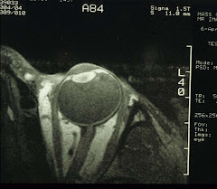
It is then possible to obtain high-resolution MR images of the eye and water-suppressed proton spectra of the vitreous. Again, for the time being.